Chapter: Human Nervous System and Sensory Organs : Spinal Cord and Spinal Nerves
Spinal Ganglion and Posterior Root - Spinal Cord

Spinal Ganglion and Posterior Root
The
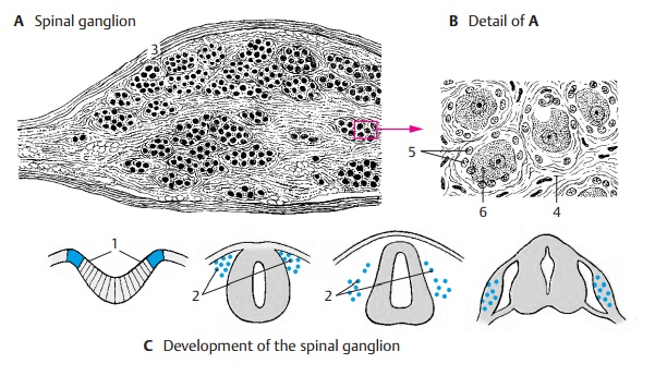
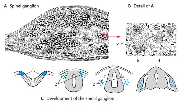
posterior spinal root contains a spindle-shaped bulge, the spinal ganglion (A), an
accumulation of cell bodies of sensory neurons; their bifurcated processes
send one branch to the periphery and the other branch to the spinal cord. They
lie as cell clusters or as cell rows between the bundles of nerve fibers.
Development of the ganglia (C).The
cellsoriginate from the lateral zone of the neuralplate (C1);
however, they do not participatein the formation of the neural tube but re-main
at both sides as the neural crest (C2). Hence, the spinal ganglia can be
regarded as gray matter of the spinal cord that became translocated to the
periphery. Other deriva-tives of the neural crest are the cells of the
autonomic ganglia, the paraganglia, and the adrenal medulla.

From the
capsule (A3) of the spinal
gan-glion, which merges into the perineurium of the spinal nerve, connective
tissue extends to the interior and forms a sheath around each neuron (endoganglionic connectivetissue) (B4). The innermost sheath, however,is
formed by ectodermal satellite cells
(BE5) and is surrounded by a basal
membrane comparable to that around the Schwann cells of the peripheral nerve.
The large nerve cells (B6, E) with
their myelinated process conglomerated into a glomerulus represent only one-third of the ganglion. They trans-mit
impulses of epicritic sensibility.
The remainder consists of medium-sized and small ganglion cells with poorly
myeli-nated or unmyelinated nerve fibers which are thought to conduct pain
signals and sen-sations from the intestine. There are also some multipolar
nerve cells.

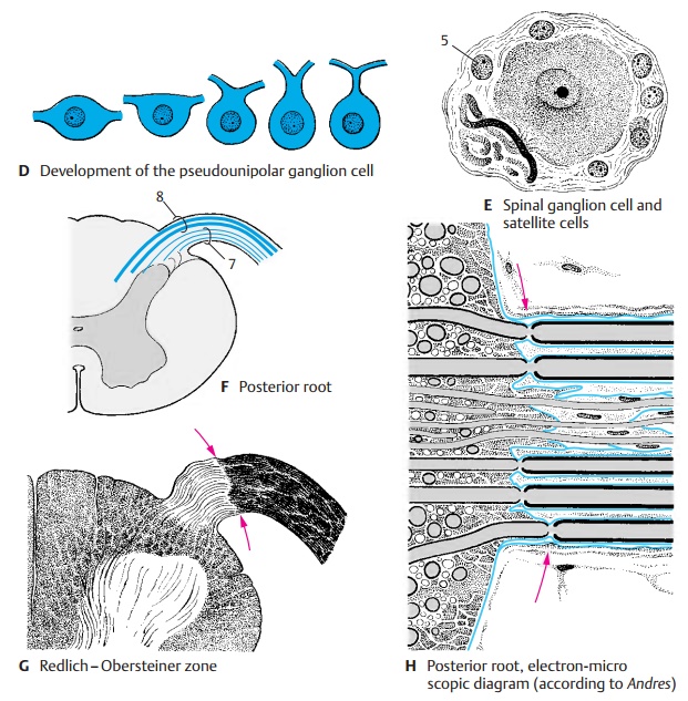
Development of the ganglion cells (D). Thespinal
ganglion cells are initially bipolarcells.
During development, however, thetwo processes fuse to form a single trunk which
then bifurcates in a T-shaped man-ner. The cells are therefore called pseudo-unipolar nerve cells.
The
posterior root is thicker than the ante-rior root. It contains fibers of
various cali-bers, two-thirds of them being poorly my-elinated or unmyelinated
fibers. The thin poorly myelinated and unmyelinated fibers, which transmit
impulses of the protopathicsensibility,
enter through the lateralpart of the root into the spinal cord (F7). The thick myelinated fibers
transmit impulses of the epicritic
sensibility and enter through the median part of the root into the spinal
cord (F8).
At the
entrance into the spinal cord, there is a narrow zone where the myelin sheaths
are very thin so that the fibers appear unmyeli-nated. This zone is regarded as
the boundarybetween the central and the
peripheral nervous systems (Redlich
– Obersteiner zone)(G). In the
electron-microscopic image (H),
however, this boundary does not exactly coincide with the Redlich – Obersteiner
zone. For each axon, the boundary is marked by the last node of Ranvier prior
to the en-trance into the spinal cord. Up to this point, the peripheral myelin
sheath is surrounded by a basal membrane
(blue in H). The next internode no
longer has a basal membrane. For unmyelinated fibers, the boundary is also
marked by the basal membrane of the enveloping Schwann cell. Thus, the basal
membranes around the spinal cord form a boundary that is only penetrated by the
axons.
Related Topics