Chapter: Human Nervous System and Sensory Organs : Spinal Cord and Spinal Nerves
Structure of Spinal Cord

The Spinal Cord
Structure
The gray matter, substantia
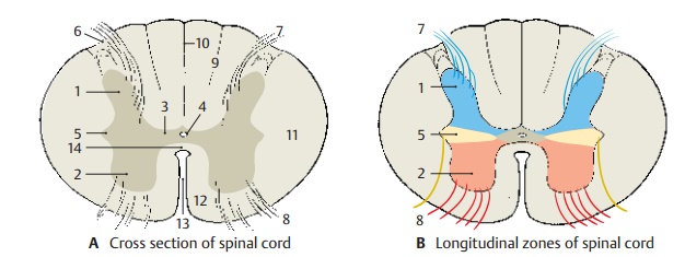
grisea (nerve cells), appears in transverse section of the spinal cord as a
butterfly configuration sur-rounded by the white
matter, substantia alba(fiber
tracts). We distinguish on either side a dorsal
horn (posterior horn) (AB1) and a ven-tral horn (anterior
horn) (AB2). Both formcolumns in
the longitudinal dimension of the spinal cord, the anterior column and the posterior
column. Between them lies thecen-tral
intermediate substance (A3) with
theobliterated central canal (A4).
In the thoracic spinal cord, the lateral
horn (AB5) is interposed between
the anterior and poste-rior horns. The lateral
posterior sulcus (A6) is the
site where the posterior root fibers (AB7)
enter. The anterior root fibers (AB8)
leave the anterior side of the spinal cord as fine bundles.

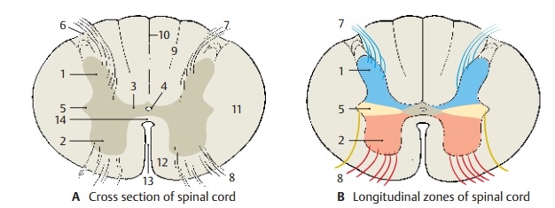
The posterior horn is derived from the alarplate (origin of
sensory neurons) and con-tains neurons of the afferent system (B). The anterior horn is derived from the basal plate (origin of
motor neurons) and contains the anterior horn cells, the efferent fibers of
which run to the muscles. The lateral horn contains autonomic nerve cells of
the sym-pathetic nervous system.
The white matter is subdivided into the dorsal column,or posterior
funiculus(A9),which reaches from
the posterior septum (A10) to the posterior horn, the lateralcolumn, orlateral funiculus(A11),
whichreaches from the posterior horn to the ante-rior root, and the ventral column, or anteriorfuniculus (A12),
which reaches from theanterior root to the anterior fissure (A13). The latter two form the anterolateral column. The white commissure (A14) connects the two halves of the spinal cord.


Related Topics