Chapter: Medicine Study Notes : Haematology and Immunology
Microscopy of Abnormal Cells
Microscopy of Abnormal Cells
Red Blood Cells
·
Oval macrocytes + hypersegmented
neutrophils Þ megaloblastic anaemia (B12/folate)
·
Target cell RBCs (haemoglobin in
the middle – non-specific): most commonly seen in patients with liver disease
(eg too much alcohol)
·
Small pale RBCs, target cells + pencil
poikilocytes (elongated RBCs) Þ iron deficiency
·
Rouleaux: red cells stack like
coins, fall fast if high ESR. Stick
together due to immunoglobulin or
·
fibrinogen). Causes: inflammation, myeloma
·
No lighter patch in middle Þ
spherocytes
·
Spherocytosis: if only some RBCs,
then spleen has taken out a bit of membrane => autoimmune haemolytic
anaemia. If all RBCs are spherocytes then hereditary spherocytosis. If
spherocytes + reticulocytes then spherocytic anaemia
·
Cygnet shape (ring form with blue
circumference) inside cell Þ malaria parasite
·
Tear drop red cells Þ
myelofibrosis or polycythaemia
·
Polychromasia Þ some red cells a bit blue due to stain – still contain some RNA
·
Reticulocytes: normal is 0.2 –
2%. Look big and blue. Will be high in anaemia (except ?anaemia of chronic
disease)
·
Howell-Jolly Bodies: Little
purple/black dots (like a ball bearing) in RBC = remnant DNA that hasn‟t been
removed by the spleen. Seen in splenectomy patients
·
Fragmented cell: red cell sliced
in circulation (DIC, artificial heart valve)
White Blood Cells
·
Normal lymphocyte: small,
little/no cytoplasm
·
Neutrophils have multilobed
nuclei, >= 6 lobes is hypersegmented (megaloblastic anaemia: B12, folate.
Also drugs, chemotherapy, renal failure)
·
Plasma cell: eccentric nuclei,
clock-face chromatin. If eccentric nucleus (clear area next to nucleus) in bone
marrow Þ multiple myeloma
· Neutrophil maturation:
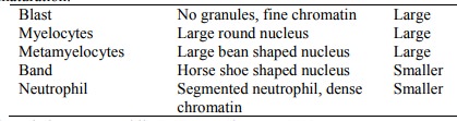
·
Normal differentiation:
Neutrophils 80%, Lymphocytes 20%
·
Lymphocyte: Toxic Changes (i.e.
„switched on‟): granules, vacuoles, Dohle bodies (blue clumps in cytoplasm), nuclear
clumping. Strong indicator of bacterial infection
·
If high lymphocytes and lots of
„atypical lymphocytes‟ then viral infection: EBV, HIV, CMV
·
Auer rods in a blast Þ acute
myeloblastic leukaemia
·
Eosinophil: normal is reddish
cytoplasmic granules. In toxoplasmosis, allergy (asthma, drugs, etc), gut parasites
·
Bone marrow biopsy: normal is
about ½ fat, ½ cellular
Leukaemias
·
Smudge cells Þ
CLL. Middle aged, significant
lymphadenopathy
·
WCC, enlarged lymph nodes, splenomegaly, lots of white cells, majority
are mature neutrophils Þ CGL (= CML)
·
Acute leukaemias: cells not
mature
Related Topics