Chapter: Medicine Study Notes : Respiratory
Lung Cancer
Lung Cancer
Smoking
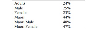
· 1998 Statistics for NZ:
· Cigarette smoking and lung cancer:
o Relative risk is 10 times in regular smoker, 20 times in those smoking > 40 per day
o Most important avoidable cause in 20 – 30% of cancers: including Respiratory tract, liver, stomach, cervix
o Tobacco and alcohol have a multiplicative relationship in oral cavity, throat and oesophagus
o Fall in lung cancer mortality begins 5 – 9 years after quitting, back to baseline at 14 years
o Abnormal cytology and squamous metaplasia in smokers
· Passive smoking:
o Passive smoking: relative risk is 3 times normal
o Relationship to URTI in children
o Possibility of younger children being affected e.g. SIDS
o Children of smokers more likely to smoke
· Active Smoking:
o Demonstrates that knowledge/education is insufficient to ensure behaviour or behaviour change
o Health promotion principles of acting at all levels (i.e. individual/community/government) to make healthy behaviour the easy choice
· Measurement: Pack-years = (cigarettes per day * years smoked) / 20
· Smoking cessation:
o Listen first: Why do you smoke? (If it‟s stress – what will you do in the future)
o What do you know about risks (don‟t assume they know about risks – maybe information lack or cognitive dissonance)
o Estimate cost for them: what would you do with $2-3,000 per year
o Give a positive message: do you want to live longer/better
o Need to negotiate with patient: be smart not paternalistic, be realistic, honest
o Always put smoking on problem list
o Information: Quit Book or Can Quit (from cancer society). Quitline 0800 778 778
Epidemiology of Lung Cancer
· Commonest cancer in the world
· In New Zealand, leading cause of cancer death in men (23%, bowel 15%, prostate 14%) and third most common in women. Maori women have the highest death rate from lung cancer of any female population in the world
· Males predominate. Females catching up
· 60% not resectable at the time of diagnosis
· 23% of all lung cancers are mixed
· Smoking:
o > 90% are caused by smoking and are therefore preventable
o 25% of lung cancer in non-smokers is due to passive smoking
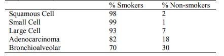
· Types according to smoking status:
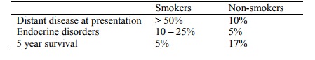
· Presentation and Survival:
· Relative incidence changing rapidly:
o ¯Squamous cell
o Adenocarcinoma (now more common than squamous cell in most countries)
o Bronchioalveolar carcinoma
o Large cell constant
Presentation
· Fatigue 84%
· Cough 71%
· Dyspnoea 59%
· Anorexia 57%
· Pain 48%
· Haemoptysis 25%
Diagnosis
· Cytology necessary for management. Use the least invasive route (eg FNA of a neck node if there is one)
· Sputum cytology
· Bronchoscopy:
o Can do washings, brushings, biopsy or lavage (to get more distal stuff)
o If can‟t produce sputum sample, can nebulise with hypo-osmotic saline to induce sputum
o 1% of transbronchial biopsy ® haemorrhage or pneumonia
· Fine Needle Aspiration (FNA): good for peripheral tumours
Types of Lung Cancer
· Squamous Cell Carcinoma:
o Most common form
o Males > females, with age
o Central tumour: presents late with invasion of lymph nodes
o Can block airway ® distal pneumonia
o Pathogenesis: BPDE in smoke binds p53 mutational hot spots ® mutation. Sequence of changes from squamous metaplasia to dysplasia to carcinoma in situ to invasive carcinoma
o Macroscopic: Arises in major bronchus, grey-white hard granular neoplasm, central cavitation in large cancers, uninvolved lung shows smoking related pathology (eg emphysema)
o Microscopic appearance: pink when stained (due to cytoplasm), keratin whirls and intracellular bridges (diagnostic), band in central cytoplasm, large irregular nucleus, nuclear pleomorphism, hyperchromatism (ie darker), coarse chromatin clumping, mitosis, large nucleoli, usually arranged in sheets
o Complications: metastatic disease to lymph nodes, brain, liver and adrenals
o Overall five year survival 10%
o Surgical treatment preferred: but may patients may have insufficient pulmonary reserve
· Small Cell Carcinoma:
o = „Oat cell‟ carcinoma
o Central Þ poor prognosis
o Very aggressive
o Treatment: chemotherapy +/- radiotherapy – not surgery as will have metastasised
o Neuroendocrine origin
o Pathogenesis: BPDE in smoke binds p53 mutational hot spots ® mutation
o Macroscopic description: perihilar and surround large bronchi. Grey-white or haemorrhagic. May be more extensive microscopically
o Microscopic appearance: small cells, scant cytoplasm (blue when stained – predominantly nuclei), ovoid, dense, hyperchromatic so nucleoli not usually seen, mitotically active, pleomorphic nuclei. Fragile ® crushed causing blue streaks
o Complications: metastatic disease to lymph nodes, brain, liver and adrenals
o Two year survival 25%
o Treatment: chemotherapy. Surgery useless unless palliative
· Large cell carcinoma:
o Undifferentiated (the „waste basket‟ category)
o Central
o White Þ desmoplastic
o Microscopic appearance: Can‟t tell cell of origin, contains giant cells, moderate amount of cytoplasm
o Quite aggressive
· Adenocarcinoma:
o Less common, Male = female
o Occurs peripherally not centrally Þ more easily respectable (unless into pleura – poor prognosis)
o Less association with smoking
o Association with previous scarring (eg Tb)
o Microscopic appearance: looks like it‟s trying to form glands, ascini, desmoplastic stroma
· Bronchioalveolar carcinoma:
o Distinctive variant of adenocarcinoma
o Slowly crawls along bronchioles
o Good 5 year survival but poor prognosis: drown in mucin
o Type of adenocarcinoma
· Carcinoid tumour:
o Low grade tumour derived from neuroendocrine cells
o Occurs younger (mean is 45) than the more frequent bronchogeneic carcinomas
o Occur in lung, bowel, other sites
o 90% Central types: 70% survive 5 years
o 10% Peripheral types: rarely metastasise
o Look like oat cell, but behave very differently. Grows by expansion rather than infiltration
· Mesothelioma:
o Primary pleural tumours, including benign and malignant (also tumours of the peritoneum, tunica vaginalis and pericardium)
o Benign mesothelioma does not produce pleural effusion and has no relationship to asbestos
o Malignant mesotheliomas arise in either visceral or parietal pleura, produce pleural effusion (can be unilateral) and are related to asbestos. Drain effusion and re-xray (looking for lumpy pleura). Do cytology on fluid. Invades lung and often other thoracic structures. Presents in 5th to 7th decade, with lag after exposure of > 20 years. Diagnosis by imaging and biopsy. Poor prognosis.
· Adenosquamous carcinoma: rarer tumour with squamous and glandular features. Aggressive, bulky, peripheral tumour
· Pancoast tumour/syndrome: lung cancer (usually squamous) in the apex extending to supraclavicular nodes and involving 8th cervical and 1st and 2nd thoracic nerves ® shoulder pain radiating in ulnar distribution. May also involve cervical sympathetic nerves and cause Horner‟s Syndrome (ipsilateral enophthalmos – sunken eye, ptosis, miosis and dry skin)
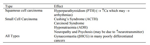
Systemic Effects of Lung Carcinoma
· Often the presenting problem:
Prognosis
· No improvement in last 40 years
· 5 year survival for all cases 13%
· Prognostic factors:
o Stage: most important factor
o Age < 40 worse (diagnosed late)
o Gender: female worse (diagnosed late)
o Site
o Size > 6 cm worse
· Staging:
o Critical to prognosis and treatment decisions
o Staging systems are regularly refined
o TNM system (not usually used for Small Cell as these have usually metastasised by diagnosis):
§ T: Size and invasion
§ N: which mediastinal nodes are involved
§ M: no metastases or metastases present
o TNMs are grouped to give stage groups ranging from IA to IV
Treatment
· Significant difference between Non-small cell and Small cell:
o Non-small Cell:
§ Resection is the gold standard, but only 20% have resectable disease at diagnosis. Surgical studies are highly selected and not representative of the general population
§ Majority will require radiotherapy. Usually palliative, (eg for haemoptysis, pain or dyspnoea) can also be radical or adjuvant
§ Chemotherapy: not used much in NZ, standard treatment in the US. Cost a factor in newer agents. Myriad of dosing regimes, combinations, etc. Can be used prior to surgery/radiotherapy to control micro-metastases/improve operability, or palliatively. Cisplatin and Etoposide are the gold standards amongst the older agents
o Small Cell:
§ 70 – 80% have metastasised at diagnosis
§ Very rapid doubling time
§ No place for surgery
§ Mainly managed with chemo +/- radiotherapy (makes a dismal outlook a bit better)
Related Topics