Chapter: Microbiology and Immunology: Antigen-Antibody Reactions
Enzyme Immunoassays - Antigen Antibody Reactions
Enzyme Immunoassays
Enzyme immunoassays (EIAs) can be used for detection of either
antigens or antibodies in serum and other body fluids of the patient. In EIA
techniques, antigen or antibody labeled with enzymes are used. Alkaline
phosphatase, horseradish peroxi-dase, and galactosidase are the enzymes used in
the EIA tests.
The commonly used EIAs are enzyme-linked immunosorbent assays
(ELISAs). The ELISA technique was first conceptualized and developed by Peter
Perlmann and Eva Engvall at Stockholm University, Sweden.
These
assays involve the use of an immunosorbent specific to either the antigen or
antibody. Following the antigen–antibody reaction, chromogenic substrate
specific to the enzyme (o-phenyl-diamine dihydrochloride for peroxidase,
p-nitrophenyl phosphate for alkaline phosphatase, etc.) is added. The reaction
is detected by reading the optical density. Usually, a standard curve based on
known concentrations of antigen or antibody is prepared from which the unknown
quantities are calculated. There are different types of ELISAs available for
the detection and quantitation of either the antigen or antibodies in serum and
other body fluids. These include: (a)
indirect ELISA, (b) sandwich ELISA, (c) competi-tive ELISA, and (d) ELISPOT assay.
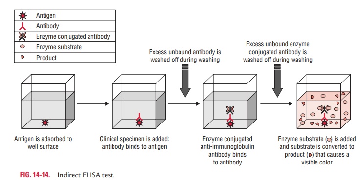
◗ Indirect ELISA
The indirect ELISA is used for the quantitative estimation of
antibodies in the serum and other body fluids. In this method, specimens are
added to microtiter plate wells coated with antigen to which specific
antibodies are to be detected. After a period of incubation, the wells are
washed. If antibody was present in the sample, antigen–antibody complex would
have
On the other hand, if the
specific antibody was not present in the specimen, there would not be any
complex formation. Next, an anti-isotype antibody conjugated with an enzyme is
added and incubated. After another washing step, a substrate for the enzyme is
added. If there was complex formation in the initial step, the second-ary
anti-isotype antibody would have bound to the primary antibody, and there would
be a chromogenic reaction between the enzyme and substrate. By measuring the
optical density val-ues of the wells, after a stop solution has been added to
arrest the chromogenic reaction, one can determine the amount of antigen–antibody
complex formed in the first step (Fig. 14-14).
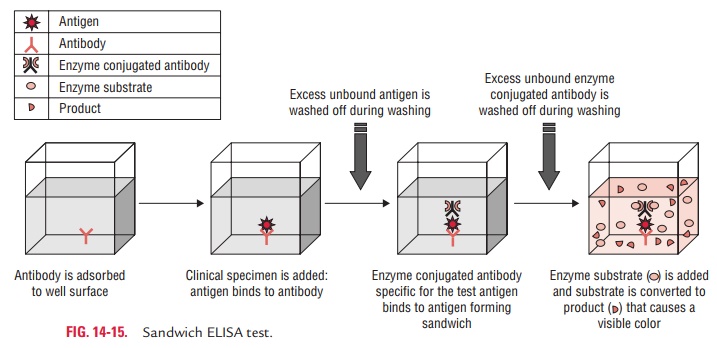
◗ Sandwich ELISA
The sandwich ELISA is used for the detection of antigen. In this
test, the known antibody is coated and immobilized onto the wells of microtiter
plates. The test sample containing the suspected antigen is added to the wells
and is allowed to react with the antibodies in the wells. After the step of
washing the well, a second enzyme-conjugated antibody specific for a dif-ferent
epitope of the antigen is added and allowed to incubate. After removing any
free secondary antibody by rewashing, the specific substrate is added, and the
ensuing chromogenic reac-tion is measured. The chromogenic reaction is then
compared with a standard curve to determine the exact amount of the antigen
present in the test sample. In a positive test, an enzyme acts on the substrate
to produce a color, and its intensity can be measured by spectrophotometer or
ELISA reader. The change of color can also be observed by the naked eye (Fig.
14-15).
◗ Competitive ELISA
Competitive ELISA is another technique used for the esti-mation of
antibodies present in a specimen, such as serum. Principle of the test is that
two specific antibodies, one conjugated with enzyme and the other present in
test serum (if serum is positive for antibodies), are used. Competition occurs
between the two antibodies for the same antigen. Appearance of color indicates
a negative test (absence of anti-bodies), while the absence of color indicates
a positive test (presence of antibodies).
In this test, the microtiter wells are coated with HIV anti-gen.
The sera to be tested are added to these wells and incu-bated at 37°C and then
washed. If antibodies are present in the test serum, antigen–antibody reaction
occurs. The antigen– antibody reaction is detected by adding
enzyme-labeled-specific HIV antibodies. In a positive test, no antigen is left
for these antibodies to act. Hence, the antibodies remain free and are washed
away during the process of washing. When substrate is added, no enzyme is
available to act on it. Therefore, positive result indicates no color reaction.
In a negative test, in which no antibodies are present in the serum, antigen in
the coated wells is available to combine with enzyme-conjugated antibodies and
the enzyme acts on the substrate to produce color.
◗ ELISPOT Assay
ELISPOT assay is a modification of ELISA. It allows the
quanti-tative determination of number of cells in a population that are producing
antibodies specific for a given antigen or an antigen for which one has a
specific antibody. These tests have found application widely in the measurement
of cytokines.
Related Topics