Chapter: Obstetric and Gynecological Nursing : Normal Pregnancy
Development of the Fertilized Ovum
Development of the Fertilized
Ovum
After fertilization the ova passes through the fallopian tube and
reaches the uterus 3 or 4 days later. Division takes place and the fertilized
ovum divides into two cells, and then into four, then eight, and sixteen and
soon until a cluster of cells is formed known as the morula.
These divisions occur quite slowly about once every 12 hours. Next,
fluid filled the cavity or blastocele appears in the morula which now becomes
known as the blastocyst.
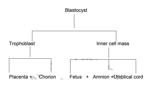
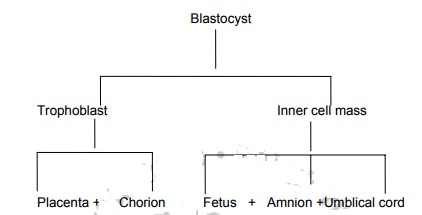
Around the out side of the blastocyst there is a single layer of cell
known as the trophoblast while the remaining cells are clumped together at one
end forming the inner cell mass. The trophoblast will form the placenta and
chorion, while the inner cell becomes the fetus, umbilical cord and the amnion.
Embedding of the blastocyst is normally completed by the 11th day after ovulation and the
endometrium closes over it completely.
The Decidua
This is the name given to the endometrium during pregnancy.
Three layers are found in decidua.
·
The basal layer lies immediately above the myometrium.
·
The functional layer consists of tortus glands which are rich in
secretions.
·
The compact layer forms the surface of the decidua and is composed of
closely packed stroma cells and the neck of the glands
The Trophoblast
Those trophoblastic cells differentiate into layers, the outer
syncitiotrophoblast (syncitium), and inner cytotrophoblast and below this, a
layer of mesoderm or primitive mesenchyme.
The syncitiotrophoblast is composed of nucleated protoplasm which is
capable of breaking down tissue as in the process of embedding.
The cytotrophoblast is a well defined single layer of cells which
produces a hormone known as human chorinic gonadotrophin (HCG).
The inner cell mass
While the trophroblast is developing into the placenta, which will
nourish the fetus, the inner cell mass is forming the fetus itself. The cells
differentiate into three layers, each of which will form particular parts of
the fetus.
·
The ectoderm mainly forms the skin and nervous system
·
The mesoderm forms bones and muscles and also the heart and blood
vessles, including those which are in placenta.
·
The endoderm forms mucous memberanes and glands. The three layers
together are known as the embryonic plate.
The amniotic
cavity-
lies on the side of the ectoderm; theyolk sac lies on the side of the endoderm
and provides nourishment for the embryo until the trophoblast is defficiaently
developed to take over.
Related Topics