Chapter: Medical Physiology: Urine Formation by the Kidneys: I. Glomerular Filtration, Renal Blood Flow, and Their Control
The Nephron Is the Functional Unit of the Kidney
The Nephron Is the Functional Unit of the Kidney
Each kidney in the human contains about 1 million nephrons, each capable of forming urine. The kidneycannot regenerate new nephrons.Therefore, with renal injury, disease, or normal aging, there is a gradual decrease in nephron number. After age 40, the number of functioning nephrons usually decreases about 10 per cent every 10 years; thus, at age 80, many people have 40 per cent fewer functioning nephrons than they did at age 40. This loss is not life threatening because adaptive changes in the remaining nephrons allow them to excrete the proper amounts of water, electrolytes, and waste products.
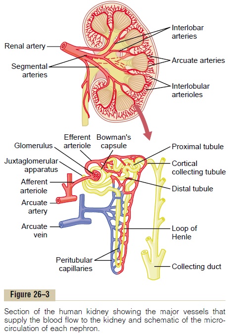
Each nephron contains (1) a tuft of glomerular cap-illaries called the glomerulus, through which large amounts of fluid are filtered from the blood, and (2) a long tubule in which the filtered fluid is converted into urine on its way to the pelvis of the kidney (see Figure 26–3).
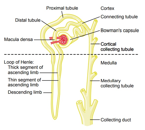
The glomerulus contains a network of branching and anastomosing glomerular capillaries that, com-pared with other capillaries, have high hydrostatic pressure (about 60 mm Hg). The glomerular capillar-ies are covered by epithelial cells, and the total glomerulus is encased in Bowman’s capsule. Fluid filtered from the glomerular capillaries flows into Bowman’s capsule and then into the proximal tubule, which lies in the cortex of the kidney (Figure 26–4).
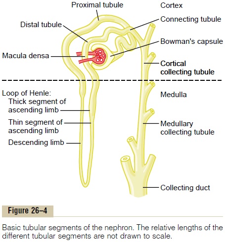
From the proximal tubule, fluid flows into the loopof Henle, which dips into the renal medulla. Each loopconsists of a descending and an ascending limb. The walls of the descending limb and the lower end of the ascending limb are very thin and therefore are called thethin segment of the loop of Henle. After the ascend-ing limb of the loop has returned partway back to the cortex, its wall becomes much thicker, and it is referred to as the thick segment of the ascending limb.
At the end of the thick ascending limb is a short segment, which is actually a plaque in its wall, known as the macula densa. As we discuss later, the macula densa plays an important role in controlling nephron function. Beyond the macula densa, fluid enters thedistal tubule, which, like the proximal tubule, lies in therenal cortex. This is followed by the connecting tubule and the cortical collecting tubule, which lead to the cor-tical collecting duct. The initial parts of 8 to 10 corticalcollecting ducts join to form a single larger collecting duct that runs downward into the medulla and becomes the medullary collecting duct. The collecting ducts merge to form progressively larger ducts that eventually empty into the renal pelvis through the tips of the renal papillae. In each kidney, there are about 250 of the very large collecting ducts, each of which collects urine from about 4000 nephrons.
Regional Differences in Nephron Structure: Cortical and Juxtamedullary Nephrons. Although each nephron has allthe components described earlier, there are some dif-ferences, depending on how deep the nephron lies within the kidney mass. Those nephrons that have glomeruli located in the outer cortex are called corti-cal nephrons; they have short loops of Henle thatpenetrate only a short distance into the medulla (Figure 26–5).
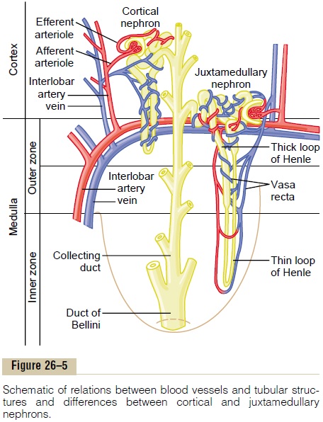
About 20 to 30 per cent of the nephrons have glomeruli that lie deep in the renal cortex near the medulla and are calledjuxtamedullary nephrons.
These nephrons have long loops of Henle that dip deeply into the medulla, in some cases all the way to the tips of the renal papillae.
The vascular structures supplying the jux-tamedullary nephrons also differ from those supplying the cortical nephrons. For the cortical nephrons, the entire tubular system is surrounded by an extensive network of peritubular capillaries. For the jux-tamedullary nephrons, long efferent arterioles extend from the glomeruli down into the outer medulla and then divide into specialized peritubular capillaries called vasa recta that extend downward into the medulla, lying side by side with the loops of Henle. Like the loops of Henle, the vasa recta return toward the cortex and empty into the cortical veins. This specialized network of capillaries in the medulla plays an essential role in the formation of a concentrated urine.
Related Topics