Chapter: 11th Zoology : Chapter 10 : Neural Control and Coordination
Sensory reception and processing
Sensory reception and processing
Our senses make us aware of changes that occur in
our surroundings and also within our body. Sensation
[awareness of the stimulus] and perception
[interpretation of the meaning of the stimulus] occur in the brain.
Receptors are classified based on their location: 1. Exteroceptors are located- at or
near the surface of the body. These are sensitive to external stimuli and
receive sensory inputs for hearing, vision, touch, taste and smell. 2. Interoceptors are located in the
visceral organs and blood vessels. They are sensitive to internal stimuli. Proprioceptors are also a kind of
-interoceptors. They provide information about position and movements of the body. These are located in the
skeletal muscles, tendons, joints, ligaments and in connective tissue coverings
of bones and muscles. Receptors based on the type of stimulus are shown in
Table 10.5.
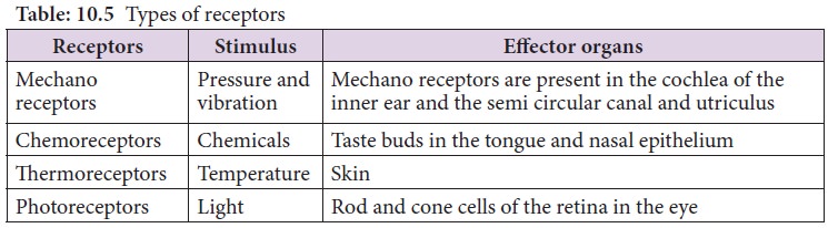
1. Photoreceptor – Eye
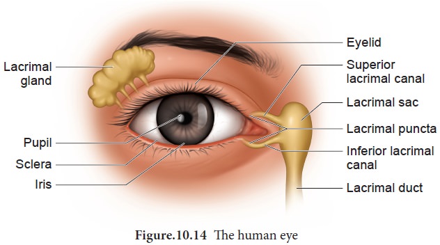
Eye is the organ of vision; located in the orbit of
the skull and held in its position with the help of six extrinsic muscles. They
are superior, inferior, lateral, median
rectus muscles, superior oblique and
inferior oblique muscles. These muscles aid in the movement of the eyes and they receive their nerve innervation from
III, IV and VI cranial nerves. Eyelids, eye lashes and eye brows are the
accessory structures useful in protecting the eyes. The eye lids protect the
eyes from excessive light and foreign objects and spread lubricating secretions
over the eye balls.
Eyelashes and the eyebrows help to protect the
eyeballs from foreign objects, perspiration and also from the direct rays of
sunlight. Sebaceous glands at the
base of the eyelashes are called ciliary
glands which secrete a lubricating fluid into the hair follicles. Lacrymal glands, located in the upper
lateral region of each orbit, secrete tears. Tears are secreted at the rate of
1mL/day and it contains salts, mucus and lysozyme
enzyme to destroy bacteria.
The conjunctiva is a thin, protective mucous
membrane found lining the outer surface of the eyeball (Figure 10.14).
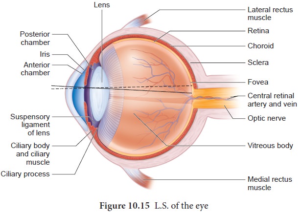
The eye has two compartments, the anterior- and posterior compartments. The anterior compartment has two chambers,
first one lies between the cornea and iris and the second one lies between the
iris and lens. These two chambers are filled with -watery
fluid called aqueous humor. The
posterior compartment lies between the lens and retina and it is filled with a
jelly like fluid called vitreous humor that helps to retain the spherical
nature of the eye. Eye lens is
transparent and biconvex, made up of long columnar epithelial cells called lens fibres. These cells are
accumulated with the proteins called crystalline.
The eye ball
The eye ball is spherical in nature. The anterior
one - sixth of the eyeball is exposed; the remaining region is fitted well into
the orbit. The wall of the eye ball consists of three layers: fibrous Sclera, vascular Choroid and sensory Retina
(Figure 10.15).
The outer coat is composed of dense non -vascular
connective tissue. It has two regions: the anterior cornea and the posterior
sclera. Cornea is a non-vascular transparent coat formed of stratified squamous
epithelium which helps thecornea to renew continuously as it is very vulnerable
to damage from dust. Sclera forms the white of the eye and protects the
eyeball. Posteriorly the sclera is innervated by the optic nerve. At the
junction of the sclera and the cornea, is a channel called ‘canal of schlemm’ which continuously drains out the excess of
aqueous humor.
![]()
Choroid is highly vascularized pigmented layer that nourishes all the eye layers and its pigments absorb light to prevent internal reflection.
Anteriorly the choroid thickens to form the ciliary body and iris. Iris is the coloured portion of the eye lying between the cornea and lens. The aperture at the centre of
the iris is the pupil through which
the light enters the inner chamber. Iris is made of two types of muscles the dilator papillae (the radial muscle) and the sphincter papillae (the circular muscle).In the bright light, the circular muscle in
the iris contract; so that the size of pupil decreases and less light enters
the eye. In dim light, the radial muscle in the iris contract; so that the
pupil size increases and more light enters the eye. Smooth muscle present in
the ciliary body is called the ciliary
muscle which alters the convexity of the lens for near and far vision. The
ability of the eyes to focus objects at varying distances is called accommodation which is achieved by suspensory ligament, ciliary muscle and ciliary body. The suspensory ligament
extends from the ciliary body and helps to
hold the lens in its upright position. The ciliary body is provided with
blood capillaries that secrete a watery fluid called aqueous humor that fills the anterior chamber.
Retina forms the
inner most layer of the eye and it contains two regions: A sheet of pigmented epithelium (non visual part)
and neural visual regions. The neural
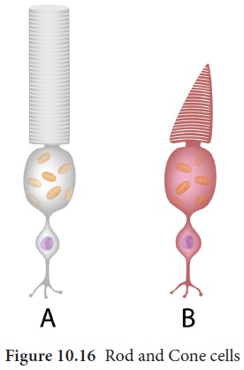
retina layer contains three types of
cells: photoreceptor cells – cones and
rods (Figure 10.16 and Table 10.6), bipolar
The yellow flat spot at the centre of the posterior
region of the retina is called macula lutea which is responsible for
sharp detailed vision. A small depression present in the centre of the yellow
spot is called fovea centralis which
contains only cones. The optic nerves and the retinal blood vessels enter the
eye slightly below the posterior pole, which is devoid of photo receptors;
hence this region is called blind spot.
Mechanism of vision
When light enters the eyes, it gets refracted by
the cornea, aqueous humor and lens and it is focused on the retina and
excites the rod and cone cells. The photo pigment consists of Opsin, the
protein part and Retinal, a
derivative of vitamin A. Light induces dissociation of retinal from opsin and
causes the structural changes in opsin. This generates an action potential in
the photoreceptor cells and is transmitted by the optic nerves to the visual
cortex of the brain, via bipolar cells, ganglia and optic nerves, for the
perception of vision.
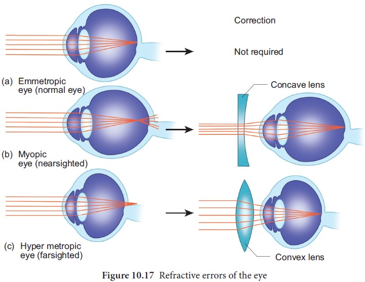
Refractive errors of eye
Myopia (near sightedness): The affected person can see the nearby objects but not the distant objects. This condition may result due to an elongated eyeball or thickened lens; so that the image of distant object is formed in front of the yellow spot. This error can be corrected using concave lens that diverge the entering light rays and focuses it on the retina.
Hypermetropia (long sightedness): the affected person can see only the distant objects clearly but not the objects nearby. This condition results due to a shortened eyeball and thin lens; so the image of closest object is converged behind the retina. This defect can be overcome by using convex lens that converge the entering light rays on the retina.
Presbyopia:
Due to
aging, the lens loses elasticity and the power of accommodation. Convex lenses
are used to correct this defect.
Astigmatism is due to the rough (irregular) -
curvature of cornea or lens. Cylindrical glasses are used
to correct this error (Figure 10.17).
Cataract:
Due to
the changes in nature of protein, the lens becomes opaque. It can be corrected
by surgical procedures.
2. Phonoreceptor
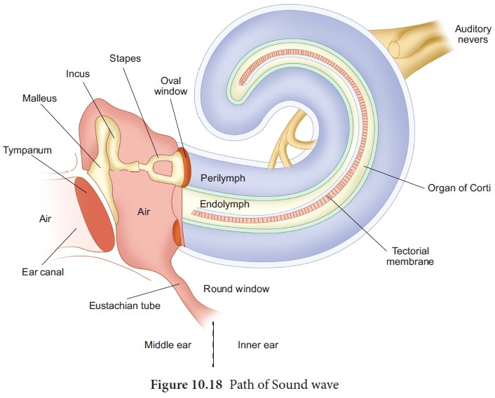
The ear is the site of reception of two senses
namely hearing and equilibrium. Anatomically, the ear is divided into three
regions: the external ear, the middle ear and internal ear.
The external ear consists of pinna, external auditory meatus and ear drum. The pinna is flap of elastic cartilage covered by skin.
It collects the sound waves. The external auditory meatus is a curved tube that
extends up to the tympanic membrane [the ear drum]. The tympanic membrane is
composed of connective tissues covered with skin outside and with mucus
membrane inside.
There are very fine hairs and wax producing
sebaceous glands called ceruminous glands in the external auditory meatus.
The combination of hair and the ear wax [cerumen]
helps in preventing dust and foreign particles from entering the ear.
The middle ear is a small air-filled cavity in the temporal bone. It is separated from the external ear by the eardrum and from the internal ear by a thin bony partition; the bony partition contains two small membrane covered openings called the oval window and the round window.
The middle ear contains three ossicles: malleus [hammer bone], incus [anvil bone] and stapes [stirrup bone] which are
attached to one another. The malleus is attached to the tympanic membrane and
its head articulates with the incus which is the intermediate bone lying
between the malleus and stapes. The stapes is attached to the oval window in
the inner ear. The ear ossicles transmit sound waves to the inner ear. A tube
called Eustachian tube connects the middle ear cavity with the pharynx. This
tube helps in equalizing the pressure of air on either sides of the ear drum.
Inner ear is the
fluid filled cavity consisting of two parts, the bony labyrinth and the
membranous labyrinths. The bony labyrinth consists of three areas: cochlea, vestibule and semicircular canals. The cochlea is a coiled portion consisting
of 3 chambers namely: scala vestibuli
and scala tympani- these two are
filled with perilymph; and the scala media is filled with endolymph. At the base of the cochlea,
the scala vestibule ends at the ‘oval window’ whereas the scala tympani ends at
the ‘round window’ of the middle ear. The chambers scala vestibuli and scala
media are separated by a membrane called Reisner’s membrane whereas the scala
media and scala tympani are separated by a membrane called Basilar membrane (Figure 10.19).
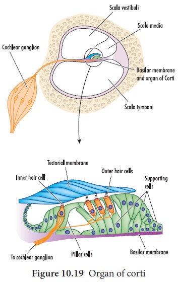
Organ of corti
The organ of
corti (figure.10.19) is a sensory ridge located on the top of the Basilar membrane and it contains numerous hair cells that are arranged in
four rows along the length of the
basilar membrane. Protruding from the apical part of each hair cell is hair
like structures known as stereocilia.
During the conduction of sound wave, stereocilia makes a contact with the stiff
gel membrane called tectorial membrane,
a roof like structure overhanging the organ of corti throughout its length.
Mechanism of hearing
Sound waves entering the external auditory meatus
fall on the tympanic membrane. This causes the ear drum to vibrate, and these
vibrations are transmitted to the oval window through the three auditory
ossicles. Since the tympanic membrane is 17-20 times larger than the oval
window,
This increased pressure
generates pressure waves in the fluid of perilymph. This pressure causes the round
window to alternately bulge outward and inward meanwhile the basilar membrane
along with the organ of Corti move up and down. These movements of the hair
alternately open and close the mechanically gated ion channels in the base of
hair cells and the action potential is propagated to the brain as sound
sensation through cochlear nerve.
Defects of Ear
Deafness may be temporary or permanent. It can be
further classified into conductive deafness and sensory-neural deafness. Possible causes for conductive deafness
may be due to
i.
the blockage of ear canal with earwax,
iii.
Middle ear infection with fluid accumulation
iii.
Restriction of ossicular movement.
In sensory
-neural deafness, the defect may be in the organ of Corti or the auditory
nerve or in the ascending auditory pathways or auditory cortex.
Organ of Equilibrium
Balance is part of a sense called proprioception,
which is the ability to sense
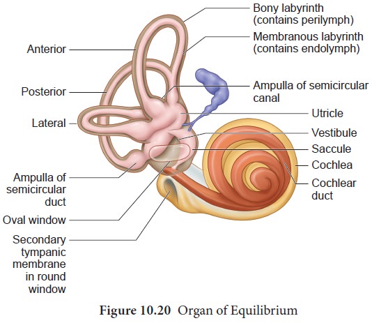
The organ of balance is known as the vestibular
system which is located in the inner ear next to the cochlea. The
vestibular system is composed of a series of fluid filled sacs and
tubules.These sacs and tubules contain endolymph and are kept in the
surrounding perilymph (Figure-10.20). These two fluids, perilymph and
endolymph, respond to the mechanical forces, during changes occurring in body
position and acceleration (Figure 10.21).
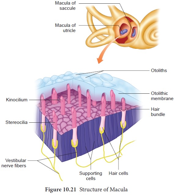
The utricle and saccule are two membranous sacs,
found nearest the cochlea and contain equilibrium receptor regions called maculae that are involved in detecting
the linear movement of the head. The maculae contain the hair cells that act as
mechanorecptors. These hair cells are embeded in a gelatinous otolithic
membrane that contains small calcareous particles called otoliths. This membrane adds weight to the top of the hair cells
and increase the inertia.
The canals that lie posterior and lateral to the
vestibule are semicircular canals; they are anterior, posterior and lateral canals oriented at right angles to
each other. At one end of each
semicircular canal, at its lower end has a swollen area called ampulla.
Each ampulla has a sensory area known as crista ampullaris which is formed of
sensory hair cells and supporting cells. The function of these canals is to
detect rotational movement of the head.
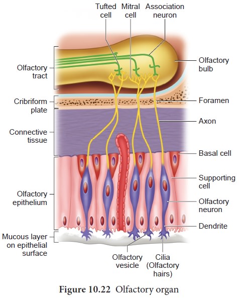
3. Olfactory receptors
The receptors for taste and smell are the
chemoreceptors. The smell receptors are excited by air borne chemicals that
dissolve in fluids. The yellow coloured patches of olfactory epithelium form
the olfactory organs (figure.10.22) that are located on the roof of the nasal
cavity. The olfactory epithelium is covered by a thin coat of mucus layer below
and olfactory glands bounded connective tissues, above. It contains three types
of cells: supporting cells, Basal cells
and millions of pin shaped olfactory
receptor cells (which are unusual
bipolar cells). The olfactory glands and the supporting cells secrete the mucus. The unmyelinated axons of the
olfactory receptor cells are gathered to form the filaments of olfactory nerve
[cranial nerve I] which synapse with cells of olfactory bulb. The impulse,
through the olfactory nerves, is transmitted to the frontal lobe of the brain
for identification of smell and the limbic system for the emotional responses
to odour.
Gustatory receptor: The sense of taste is considered to be the most pleasurable of all senses. The tongue is provided with many small projections called papillae which give the tongue an abrasive feel. Taste buds are located mainly on the papillae which are scattered over the entire tongue surface.
Most taste buds are seen on the tongue (Figure
10.23) few are scattered on the soft palate, inner surface of the cheeks,
pharynx and epiglottis of the larynx. Taste buds are flask-shaped and consist
of 50 – 100 epithelial cells of two major types.
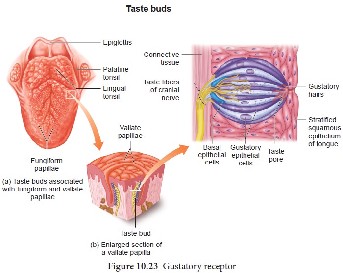
Gustatory
epithelial cells (taste cells) and Basal epithelial cells (Repairing cells) Long microvilli called gustatory hairs project from the tip of
the gustatory cells and extends through a taste pore to the surface of the
epithelium where they are bathed by saliva. Gustatory- hairs are the sensitive
portion of the gustatory cells and they have sensory dendrites which send the
signal to the brain. The basal cells that act as stem cells, divide- and
differentiate into new gustatory cells (Figure 10.23).
Skin-Sense of touch
Skin is the sensory organ of touch and is also the
largest sense organ. This sensation comes from millions of microscopic sensory
receptors located all over the skin and associated with the general sensations
of contact, pressure, heat, cold and pain. Some parts of the body, such as the
finger tips have a large number of these receptors, making them more sensitive.
Some of the sensory receptors present in the skin (Figure 10.24) are:

•
Tactile
merkel disc is light touch receptor lying in the deeper layer
of epidermis.
•
Hair
follicle receptors are light touch receptors lying around the hair follicles.
•
Meissner’s
corpuscles are small light pressure receptors found just
beneath the epidermis in the dermal
papillae. They are numerous in hairless skin areas such as finger tips and
soles of the feet.
•
Pacinian
corpuscles are the large egg shaped receptors found scattered deep in the dermis and monitoring
vibration due to pressure. It allows to detect different textures, temperature,
hardness and pain
•
Ruffini
endings which lie in the dermis responds-
to continuous pressure.
• Krause end bulbs are thermoreceptors that sense temperature.
Melanocytes are the cells responsible for producing
the skin pigment, melanin, which gives skin its colour and protects it from the
sun's UV rays. Vitiligo (Leucoderma) is a condition in which the melanin
pigment is lost from areas of the skin, causing white patches, often with no
clear cause. Vitiligo is not contagious. It can affect people of any age,
gender, or ethnic group. The patches appear when melanocytes fails to synthesis
melanin pigment.
Related Topics