Chapter: 11th Zoology : Chapter 10 : Neural Control and Coordination
Reflex action and Reflexarc
Reflex action and Reflexarc
When dust falls in our eyes, the eyelids close
immediately not waiting for our willingness; on touching a hot pan, the hand is
withdrawn rapidly. Do you know how this happens?
The spinal cord remains as a connecting functional
nervous structure in between the brain and effector organs. But sometimes when
a very quick response is needed, the spinal cord can effect motor
initiation as the brain and brings about an effect. This rapid action by spinal
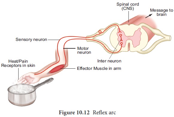
cord is called reflex action. It is a fast, involuntary, unplanned sequence of
actions that occurs in response to a particular stimulus. The nervous elements
involved in carrying out the reflex action constitute a reflex arc or in other
words the pathway followed by a nerve impulse to produce a reflex action is
called a reflex arc -(Figure 10.12).

Functional components of a reflex arc
Sensory Receptor - It is a sensory structure that
responds to a specific stimulus. Sensory Neuron - This neuron takes the sensory
impulse to the grey (afferent) matter of the spinal cord through the dorsal
root of the spinal cord.
Interneurons - One or two interneurons may serve to
transmit the impulses from the sensory neuron to the motor neuron.
Motor Neuron - it transmits impulse from CNS to the
effector organ.
Effector Organs -It may be a muscle or gland which
responds to the impulse received. There are two types of reflexes. They are
1. Unconditional reflex is an inborn reflex for an unconditioned stimulus. It does not need any past experience, knowledge or training to occur; Ex: blinking of an eye when a dust particle about to fall into it, sneezing and coughing due to foreign particle entering the nose or larynx.
1.
Conditioned
reflex is a respone to a stimulus that has been acquired by learning. This does not naturally exists in animals.
Only an experience makes it a part of the behaviour. Example: excitement of
salivary gland on seeing and smelling a food. The conditioned reflex was first
demonstrated by the Russian physiologist Pavlov
in his classical conditioning experiment in a dog. The
cerebral cortex controls the conditioned reflex.
Peripheral Neural System (PNS)
PNS consists of all nervous tissue outside the CNS.
Components of PNS include nerves, ganglia, enteric plexuses and sensory
receptors. A nerve is a chord like structure that encloses several neurons
inside. Ganglia (singular-ganglion) are small masses of nervous tissue,
consisting primarily of neuron cell bodies and are located outside the brain
and spinal cord. Enteric plexuses are extensive networks of neurons located in
the walls of organs of the gastrointestinal tract. The neurons of these
plexuses help in regulating the -digestive system. The specialized structure
that helps to respond to changes in the environment i.e. stimuli are called sensory receptor which triggers nerve impulses along the afferent fibres to
CNS. PNS comprises of cranial nerves
arising- from the brain and spinal- nerves arising from the spinal cord.
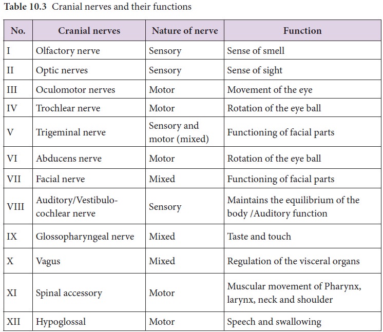
(A) Cranial
nerves: There are 12 pairs of cranial nerves, of which the first two pairs arise from the fore brain and the remaining
10 pairs from the mid brain. Other than the Vagus nerve, which extends into the
abdomen, all cranial nerves serve the head and face (Table 10.3).

(B) Spinal
nerves: 31 pairs of spinal nerves emerge out from the spinal cord through spaces called the intervertebral foramina found between the adjacent vertebrae. The
spinal nerves are named according to the region of vertebral column from which
they originate
i. Cervical nerves (8 pairs)
ii. Thoracic nerves (12 pairs)
iii. Lumbar nerves (5 pairs)
iv. Sacral nerves (5 pairs)
v. Coccygeal nerves (1 pair)
Each spinal nerve is a mixed nerve containing both
afferent (sensory) and efferent (motor) fibres. It originates as two roots:1) a posterior dorsal root with a
ganglion outside the spinal cord and 2) an anterior ventral root with no
external ganglion.
Somatic neural
system (SNS)
The somatic
neural system (SNS or voluntary neural system) is the part of the
peripheral neural system associated with the voluntary control of body
movements via skeletal muscles. The sensory and motor nerves that innervate
striated muscles form the somatic neural system. Major functions of the somatic
neural system include voluntary movement of the muscles and organs, and reflex
movements.
Autonomic Neural System
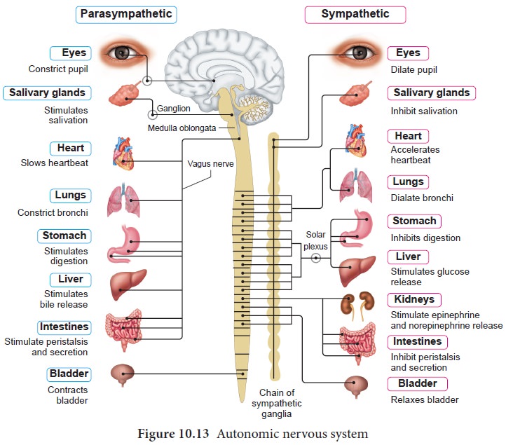
The autonomic neural system is auto functioning and
self governed. It is a part of peripheral neural system that innervates smooth
muscles, glands and cardiac muscle. This system controls and coordinates the
involuntary activities of various organs. ANS (Figure.10.13) controlling centre
is in the hypothalamus. Autonomic neural system comprises the following
components:
Preganglionic
neuron whose cell body is in the brain or spinal cord; its myelinated axon exits the CNS as part of cranial
or spinal nerve and ends in an autonomic ganglion.
Autonomic
ganglion consists of axon of preganglionic neuron and cell bodies of postganglionic neuron.
Postganglionic
neuron conveys nerve impulses from autonomic ganglia to visceral effector organs.
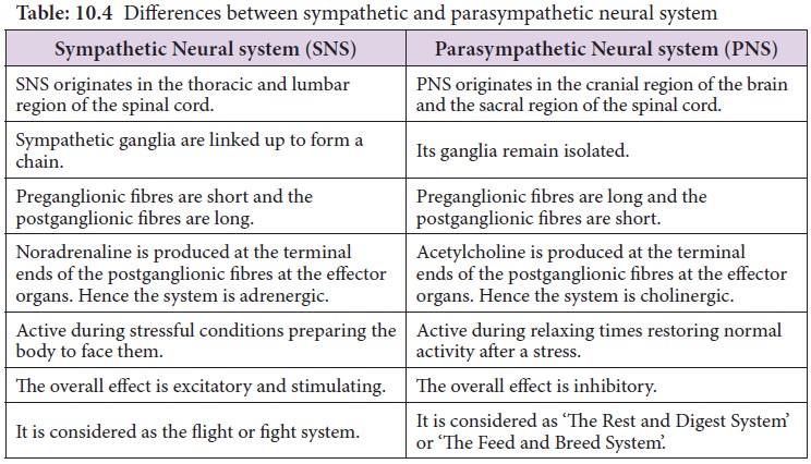
The autonomic neural system consists of Sympathetic neural system and Parasympathetic neural system (Table
10.4).
Table: 10.4 Differences between sympathetic and parasympathetic neural system

Related Topics
