Chapter: Medical Physiology: Urine Formation by the Kidneys: I. Glomerular Filtration, Renal Blood Flow, and Their Control
Physiologic Anatomy and Nervous Connections of the Bladder
Physiologic Anatomy and Nervous Connections of the Bladder
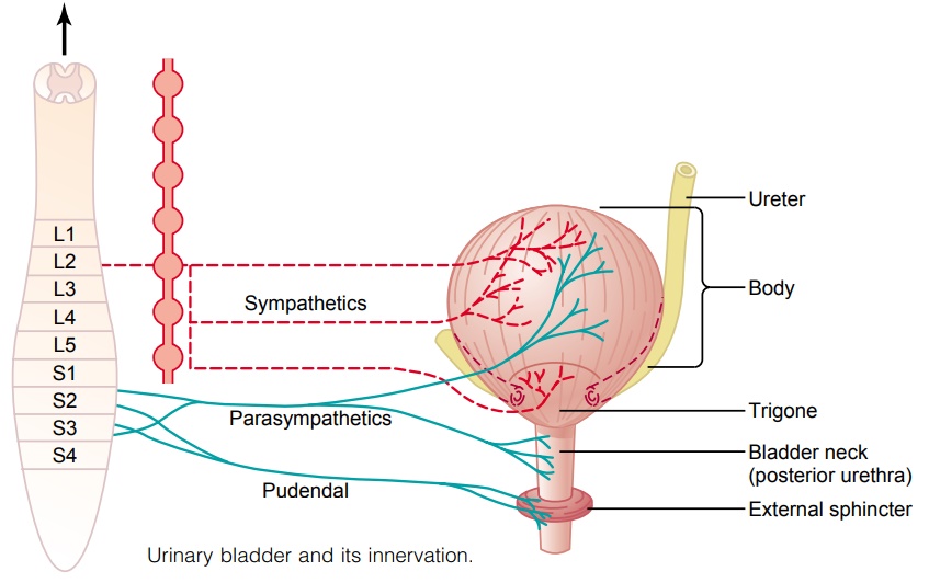
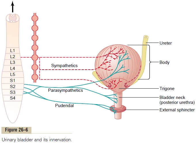
The urinary bladder, shown in Figure 26–6, is a smooth muscle chamber composed of two main parts: (1) the body, which is the major part of the bladder in which urine collects, and (2) the neck, which is a funnel-shaped extension of the body, passing inferiorly and anteriorly into the urogenital triangle and connecting with the urethra. The lower part of the bladder neck is also called the posterior urethra because of its relation to the urethra.
The smooth muscle of the bladder is called the detru- sor muscle. Its muscle fibers extend in all directions and, when contracted, can increase the pressure in the bladder to 40 to 60 mm Hg. Thus,contraction of the detrusor muscle is a major step in emptying the bladder. Smooth muscle cells of the detrusor muscle fuse with one another so that low-resistance electrical pathways exist from one muscle cell to the other. Therefore, an action potential can spread throughout the detrusor muscle, from one muscle cell to the next, to cause con- traction of the entire bladder at once.
On the posterior wall of the bladder, lying immedi- ately above the bladder neck, is a small triangular area called the trigone. At the lowermost apex of the trigone, the bladder neck opens into the posterior urethra, and the two ureters enter the bladder at the uppermost angles of the trigone. The trigone can be identified by the fact that its mucosa, the inner lining of the bladder, is smooth, in contrast to the remaining bladder mucosa, which is folded to form rugae. Each ureter, as it enters the bladder, courses obliquely through the detrusor muscle and then passes another 1 to 2 centimeters beneath the bladder mucosa before emptying into the bladder.
The bladder neck (posterior urethra) is 2 to 3 cen-timeters long, and its wall is composed of detrusor muscle interlaced with a large amount of elastic tissue. The muscle in this area is called the internal sphincter.Its natural tone normally keeps the bladder neck and posterior urethra empty of urine and, therefore, pre-vents emptying of the bladder until the pressure in the main part of the bladder rises above a critical threshold.
Beyond the posterior urethra, the urethra passes through the urogenital diaphragm, which contains a layer of muscle called the external sphincter of the bladder. This muscle is a voluntary skeletal muscle, in contrast to the muscle of the bladder body and bladder neck, which is entirely smooth muscle. The external sphincter muscle is under voluntary control of the nervous system and can be used to consciously prevent urination even when involuntary controls are attempt-ing to empty the bladder.
Innervation of the Bladder
The principal nerve supply of the bladder is by way of the pelvic nerves, which connect with the spinal cord through the sacral plexus, mainly connecting with cord segments S-2 and S-3. Coursing through the pelvic nerves are both sensory nerve fibers and motor nervefibers. The sensory fibers detect the degree of stretch inthe bladder wall. Stretch signals from the posterior urethra are especially strong and are mainly re-sponsible for initiating the reflexes that cause bladder emptying.
The motor nerves transmitted in the pelvic nerves are parasympathetic fibers. These terminate on ganglioncells located in the wall of the bladder. Short postgan-glionic nerves then innervate the detrusor muscle.
In addition to the pelvic nerves, two other types of innervation are important in bladder function. Most important are the skeletal motor fibers transmitted through the pudendal nerve to the external bladder sphincter. These are somatic nerve fibers that innervate and control the voluntary skeletal muscle of the sphinc-ter. Also, the bladder receives sympathetic innervation from the sympathetic chain through thehypogastricnerves, connecting mainly with the L-2 segment of thespinal cord. These sympathetic fibers stimulate mainly the blood vessels and have little to do with bladder con-traction. Some sensory nerve fibers also pass by way of the sympathetic nerves and may be important in the sensation of fullness and, in some instances, pain.
Related Topics