Chapter: Biochemistry: Nucleic Acids: How Structure Conveys Information
How does prokaryotic DNA supercoil into its tertiary structure?
How does prokaryotic DNA
supercoil into its tertiary structure?
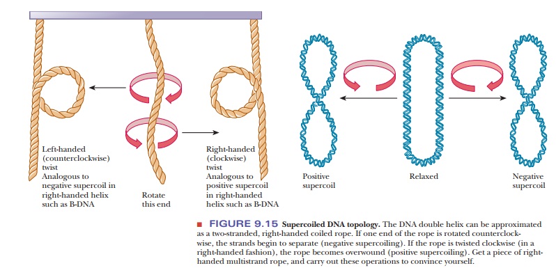
The first example of supercoiling we shall consider is the case of prokaryotic DNA. If the sugar–phosphate backbone of a prokaryotic DNA forms a covalently bonded circle, the structure is still relaxed. Some extra twists are added if the DNA is unwound slightly before the ends are joined to form the circle. A strain is introduced in the molecular structure, and the DNA assumes a new conformation to compensate for the unwinding. If, because of unwinding, a right-handed double helix acquires an extra left-handed helical twist (a supercoil), the circular DNA is said to be negatively supercoiled (Figure 9.15). Under different conditions, it is possible to form a right-handed, or positively supercoiled, structure in which there is overwinding of the closed-circle double helix. The difference between the positively and negatively supercoiled forms lies in their right- and left-handed natures, which, in turn, depend on the overwinding or underwinding of the double helix.
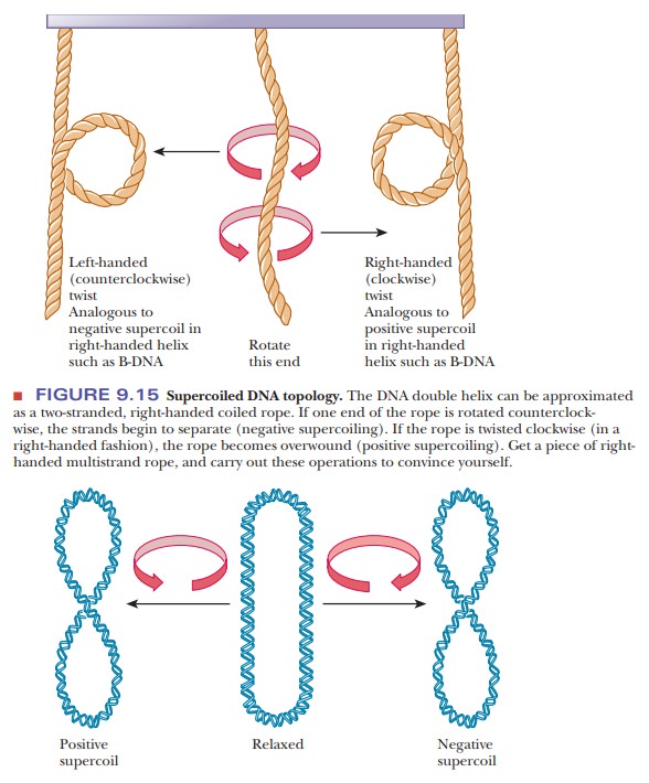
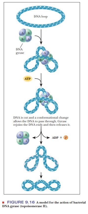
Enzymes that affect the supercoiling of DNA have been isolated from a vari-ety of organisms. Naturally occurring circular DNA is negatively supercoiled except during replication, when it becomes positively supercoiled. Cellular regulation of this process is critical. Enzymes that are involved in changing the supercoiled state of DNA are called topoisomerases, and they fall into two classes. Class I topoisomerases cut the phosphodiester backbone of one strand of DNA, pass the other end through, and then reseal the backbone. Class II topoisomerases cut both strands of DNA, pass some of the remaining DNA helix between the cut ends, and then reseal. In either case, supercoils can beadded or removed. As we shall see in upcoming, these enzymes play an important role in replication and transcription, where separation of the helix strands causes supercoiling. DNA gyrase is a bacterial topoisomerase that introduces negative supercoils into DNA. The mechanism is shown in Figure 9.16. The enzyme is a tetramer. It cuts both strands of DNA, so it is a class II topoisomerase.
Supercoiling has been observed experimentally in naturally occurring DNA. Particularly strong evidence has come from electron micrographs that clearly show coiled structures in circular DNA from a number of different sources, including bacteria, viruses, mitochondria, and chloroplasts. Ultracentrifugation can be used to detect supercoiled DNA because it sediments more rapidly than the relaxed form.
Scientists have known for some time that prokaryotic DNA is normally circular, but supercoiling is a relatively recent subject of research. Computer modeling has helped scientists visualize many aspects of the twisting and knotting of supercoiled DNA by obtaining “stop-action” images of very fast changes.
Related Topics