Chapter: Essentials of Anatomy and Physiology: Senses
Hearing - Ear
Hearing
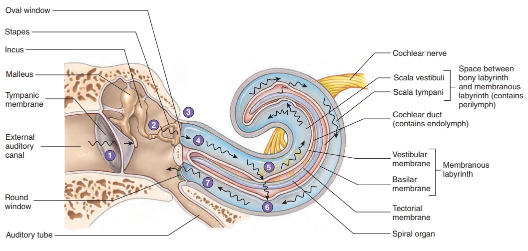
Vibrations create sound waves. Sound waves are collected by the auricle and conducted through the external auditory canal toward the tympanic membrane. Sound waves strike the tympanic mem-brane and cause it to vibrate. This vibration causes vibration of the three ossicles of the middle ear, and by this mechanical linkage, the force of vibration is amplified and transferred to the oval win-dow (figure 9.19, steps 1–3).
Vibrations of the base of the stapes, seated in the oval window, produce waves in the perilymph of the cochlea. The two scalae can be thought of as a continuous, U-shaped tube, with the oval window at one end of the scala vestibuli and the round window at the other end of the scala tympani. The vibrations of the stapes in the oval window cause movement of the perilymph, which pushes against the membrane covering the round window (figure 9.19 step 4). This phenomenon is similar to pushing against a rubber diaphragm on one end of a fluid-filled glass tube. If the tube has a rubber dia-phragm on each end, the fluid can move. If one end of the glass tube or of the cochlear tubes were solid, no fluid movement would occur.
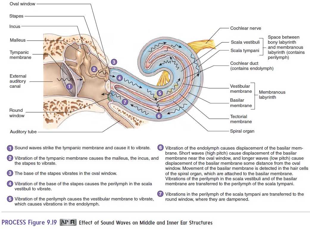
The waves produced in the perilymph pass through the vestibu-lar membrane and cause vibrations of the endolymph. Waves in the endolymph, within the cochlear duct, cause displacement of the basilar membrane. As the basilar membrane is displaced, the hair cells, seated on the basilar membrane, move with the movements of the membrane. The microvilli of the hair cells are embedded in the tectorial membrane, which is a rigid shelf that does not move. Because one end of the microvilli moves with the hair cells and their other ends are embedded in the nonmoving tectorial membrane, the microvilli bend. The bending of the microvilli stimulates the hair cells, which induces action potentials in the cochlear nerves (figure 9.19, steps 5–6).
The basilar membrane is not uniform throughout its length. The membrane is narrower and denser near the oval window and wider and less dense near the tip of the cochlea. The various regions of the membrane can be compared to the strings in a piano (i.e., some are short and thick, and others are longer and thinner). As a result of this organization, sounds with higher pitches cause maximum distortion of the basilar membrane nearer the oval window, whereas sounds with lower pitches cause maximum distortion nearer the apex of the cochlea. In each case, different hair cells are stimulated, and because of the differences in which hair cells are maximally stimulated, a person is able to detect variations in pitch. Sound volume is a function of sound wave amplitude, which causes the basilar membrane to distort more intensely and the hair cells to be stimulated more strongly.
Hearing impairment can have many causes. In general, there are two categories of hearing impairment: conduction deafness and sensorineural hearing loss (see the Diseases and Disorders table). Conduction deafness results from mechanical deficiencies—for example, destruction of the ligament that holds the malleus and incus together. Sensorineural hearing loss is caused by deficiencies in the spiral organ or nerves;for example, loud sounds can damage the delicate microvilli of the hair cells, leading to destruction of the spiral organ.
Related Topics