Chapter: Medical Physiology: Introduction to Physiology: The Cell and Its Functions
Ameboid Movement
Ameboid Movement
Ameboid movement is movement of an entire cell in relation to its surroundings, such as movement of white blood cells through tissues. It receives its name from the fact that amebae move in this manner and have pro-vided an excellent tool for studying the phenomenon.
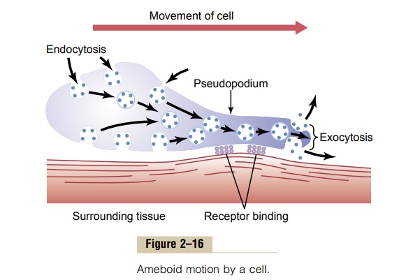
Typically, ameboid locomotion begins with protrusion of a pseudopodium from one end of the cell. The pseudopodium projects far out, away from the cell body, and partially secures itself in a new tissue area. Then the remainder of the cell is pulled toward the pseudopodium. Figure 2–16 demonstrates this process, showing an elongated cell, the right-hand end of which is a protruding pseudopodium. The membrane of this end of the cell is continually moving forward, and the membrane at the left-hand end of the cell is continually following along as the cell moves.
Mechanism of Ameboid Locomotion. Figure 2–16 shows thegeneral principle of ameboid motion. Basically, it results from continual formation of new cell membrane at the leading edge of the pseudopodium and continual absorption of the membrane in mid and rear portions of the cell. Also, two other effects are essential for forward movement of the cell. The first effect is attach-ment of the pseudopodium to surrounding tissues so that it becomes fixed in its leading position, while the
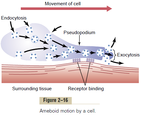
remainder of the cell body is pulled forward toward the point of attachment. This attachment is effected by receptor proteins that line the insides of exocytotic vesi-cles. When the vesicles become part of the pseudopodial membrane, they open so that their insides evert to the outside, and the receptors now protrude to the outside and attach to ligands in the surrounding tissues.
At the opposite end of the cell, the receptors pull away from their ligands and form new endocytotic vesi-cles. Then, inside the cell, these vesicles stream toward the pseudopodial end of the cell, where they are used to form still new membrane for the pseudopodium.
The second essential effect for locomotion is to provide the energy required to pull the cell body in the direction of the pseudopodium. Experiments suggest the following as an explanation: In the cytoplasm of all cells is a moderate to large amount of the protein actin.Much of the actin is in the form of single molecules that do not provide any motive power; however, these poly-merize to form a filamentous network, and the network contracts when it binds with an actin-binding protein such as myosin. The whole process is energized by the high-energy compound ATP. This is what happens in the pseudopodium of a moving cell, where such a network of actin filaments forms anew inside the enlarging pseudopodium. Contraction also occurs in the ectoplasm of the cell body, where a preexisting actin network is already present beneath the cell membrane.
Types of Cells That Exhibit Ameboid Locomotion. The mostcommon cells to exhibit ameboid locomotion in the human body are the white blood cells when they move out of the blood into the tissues in the form of tissuemacrophages. Other types of cells can also move byameboid locomotion under certain circumstances. For instance, fibroblasts move into a damaged area to help repair the damage, and even the germinal cells of the skin, though ordinarily completely sessile cells, move toward a cut area to repair the rent. Finally, cell loco-motion is especially important in development of the embryo and fetus after fertilization of an ovum. For instance, embryonic cells often must migrate long dis-tances from their sites of origin to new areas during development of special structures.
Control of Ameboid Locomotion—Chemotaxis. The mostimportant initiator of ameboid locomotion is the process calledchemotaxis. This results from the appear-ance of certain chemical substances in the tissues. Any chemical substance that causes chemotaxis to occur is called a chemotactic substance. Most cells that exhibit ameboid locomotion move toward the source of a chemotactic substance—that is, from an area of lower concentration toward an area of higher con-centration—which is called positive chemotaxis. Some cells move away from the source, which is called nega-tive chemotaxis.
But how does chemotaxis control the direction of ameboid locomotion? Although the answer is not certain, it is known that the side of the cell most exposed to the chemotactic substance develops membrane changes that cause pseudopodial protrusion.
Related Topics