Chapter: Essentials of Anatomy and Physiology: Senses
Accessory Structures of the Eye
Accessory Structures of the Eye
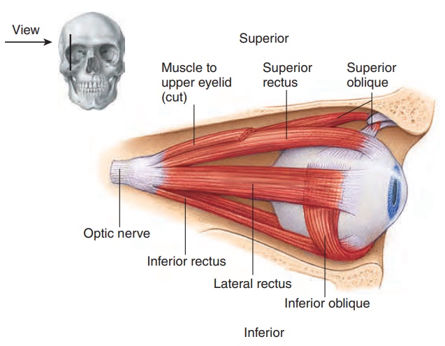
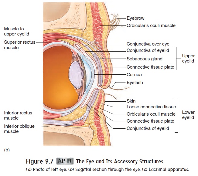
Accessory structures protect, lubricate, and move the eye. They include the eyebrows, eyelids, conjunctiva, lacrimal apparatus, and extrinsic eye muscles (figures 9.7 and 9.8).

Eyebrows
The eyebrows protect the eyes by preventing perspiration from running down the forehead and into the eyes, causing irritation. They also help shade the eyes from direct sunlight (figure 9.7a).
Eyelids
The eyelids, with their associated lashes, protect the eyes from foreign objects (figure 9.7a,b). If an object suddenly approaches the eye, the eyelids protect the eye by closing and then opening quite rapidly (blink reflex). Blinking, which normally occurs about 20 times per minute, also helps keep the eyes lubricated by spreading tears over the surface.
Conjunctiva
The conjunctiva (kon-jŭ nk-t ̄ı ′ v̆a ) is a thin, transparent mucous membrane covering the inner surface of the eyelids and the ante-rior surface of the eye (figure 9.7b). The secretions of the con-junctiva help lubricate the surface of the eye. Conjunctivitis is an inflammation of the conjunctiva (see the Diseases and Disorders table).
Lacrimal Apparatus
The lacrimal (lak′ ri-mă l; tear) apparatus consists of a lacrimal gland situated in the superior lateral corner of the orbit and a nasolacrimal duct and associated structures in the inferior medial corner of the orbit (figure 9.7c). The lacrimal gland produces tears, which pass over the anterior surface of the eye. Most of the fluid produced by the lacrimal glands evaporates from the surface of the eye, but excess tears are collected in the medial angle of the eyes by small ducts called lacrimal canaliculi (kan-̆a -lik′ ̄u -l̄ı ; lit-tle canals). These canaliculi open into a lacrimal sac, an enlarge-ment of the nasolacrimal (n̄a -z̄o -lak′ ri-m̆a l) duct, which opens into the nasal cavity. Tears lubricate and cleanse the eye. They also contain an enzyme that helps combat eye infections.
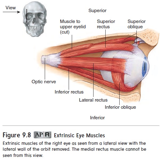
Extrinsic Eye Muscles
Movement of each eyeball is accomplished by six skeletal muscles called the extrinsic eye muscles (figure 9.8). Four of these muscles run more or less straight from their origins in the posterior portion of the orbit to their insertion sites on the eye, to attach to the four quad-rants of the eyeball. They are the superior, inferior, medial, and lat-eral rectus muscles. Two muscles, the superior and inferior obliquemuscles,are located at an angle to the long axis of the eyeball.
Related Topics