Chapter: Human Nervous System and Sensory Organs : The Eye
Visual Pathway - Eye

Visual Pathway
The
visual pathway consists of four neurons connected in tandem:
! 1st
neuron, the photoreceptors
!
2nd neuron, the bipolar
neurons of the ret-ina, which transmit the impulses from the rods and cones
to the large ganglion cells of the retina
!
3rd neuron, the large
ganglion cells, the axons of which combine to form the optic nerve and
extend to the primary visual centers (lateral
geniculate nucleus)
!
4th neuron, the geniculate
cells, the axons of which project as the optic radiation to the visual
cortex (striate area)
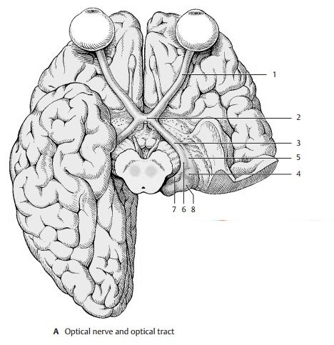
The optic nerve (A1) enters the cranial cavity through the optic canal. At the base
of the diencephalon, together with the con-tralateral optic nerve, it forms the
optic chi-asm(A2). The fiber bundle starting from thechiasm is known as the optic tract (A3). The two tracts run around the cerebral peduncles to the two
lateral geniculate bod-ies (A4).
Before reaching these, each tract divides into a lateral root (A5) and a medialroot (A6). Whereas most of the fibers runthrough the lateral root to the
lateral geniculate body, the medial fibers continue below the medial geniculate
body (A7) to the superior colliculi.
They contain visual reflex pathways. The optic nerve fibers are thought to give
off collater-als to the pulvinar of the thalamus (A8) prior to terminating in the lateral geniculate body. The optic
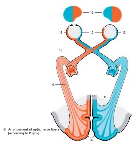
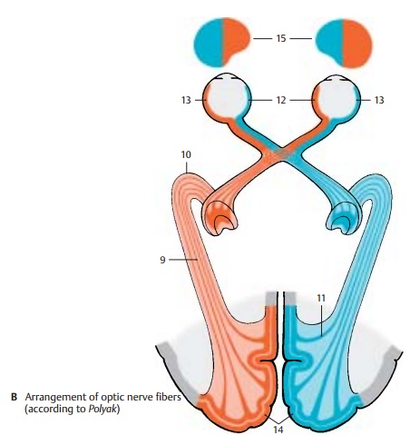
radiation (radiation of Gra-tiolet) (B9) begins at thelateral geniculatebody and extends as a broad fiber plate tothe
calcarine sulcus at the medial aspect of the occipital lobe and, while doing
so, forms the outward-arching temporal
genu (B10) . Numerous fibers
bend ros-trally (occipital genu) (B11) in the occipital lobe to reach the
anterior areas of the visual cortex.

The optic
nerve fibers originating from the nasal halves (B12) of the retina cross in theoptic chiasm. The fibers from the
temporal halves (B13) do not cross
but continue on the ipsilateral side. Hence, the right tract contains the
fibers from the temporal half of the right eye and from the nasal half of the left eye. The left
tract contains fibers from the temporal half of the left eye and from the nasal
half of the right eye. In a cross section of the tract, the crossed fibers lie
mostly ventrolaterally, and the uncrossed fibers dorsomedially; in between, the
fibers are mixed.
The crossed and uncrossed fibers
of the optic tract extend to different cell layers of the lateral geniculate
body. The number of geniculate cells, approximately one million, corresponds to
the number of optic nerve fibers. However, the optic nerve fibers usually end
on five to six cells located in different cell layers. Cor-ticofugal fibers of
the occipital cortex also end in the lateral geniculate body. They probably
control the input of impulses, as suggested by the pres-ence of axo-axonal
synapses characteristic for presynaptic inhibition.

The axons of the geniculate cells
form the optic radiation. Its fibers are arranged according to the different
regions of the retina. The fibers for the lower half of the retina, especially
those for the periphery of the retina, arch most rostrally in the temporal
genu. The fibers for the upper half of the retina and for the central region of
the mac-ula arch only slightly in the temporal lobe.
In the striate area (B14) of
the right hemi-sphere terminate the fibers for the right halves of the retinae;
hence, it receives sensory input from the left halves of the visual fields. In
the striate area of the left hemisphere there terminate the fibers of the left
halves of the retinae with input from the right halves of the visual fields.
The right hand and the right visual field are therefore both represented in the
left hemisphere, which dominates in right-handed persons.
B15
Visual fields.
Related Topics