Chapter: Human Nervous System and Sensory Organs : The Eye
Eyeball: Development, Structure
The Eyeball
Development (A)
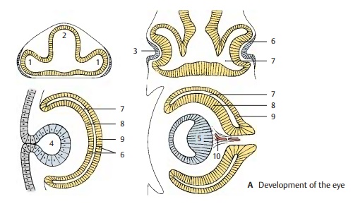
The light-sensitive part of the eye is a derivative of the diencephalon. At end of the first month of embryonic development, the two optic vesicles (A1) are formed as evagi-nations of the prosencephalon (A2). The optic vesicles then induce thickenings in the ectoderm of the head, the lens placodes(A3), which later invaginate as lens vesicles (A4). The epithelial cells of the posterior vesicle wall elongate into lens fibers (A5), which later form the main part of the lens. The cells of the anterior vesicle wall persist as lens epithelium. The anterior and posterior walls of the optic vesicle approximate each other to form the optic cup (A6). The lumen of the vesicle, originally a part of the ventricular system, the optic ventricle (A7), becomes a narrow cavity. The optic cup con-sists of an inner layer, the neural layer (A8), and an outer layer, the pigmented layer (A9); both are layers of the retina. Thehyaloidartery (A10) first extends to the lens butlater regresses.
Structure (B)
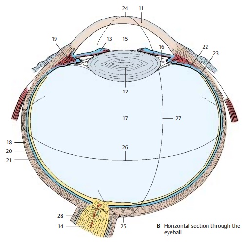
The anterior aspect of the eyeball consists of the transparent cornea (B11). Behind it lies the lens of the eye (B12), overlaid by the iris (B13) with its central opening, the pupil. The optic nerve (B14) exits at the posterior wall ofthe eyeball, slightly medial to the optic axis. There are three cavities in the eye:
! The anterior chamber (B15), which is bordered by cornea, iris, and lens
! The posterior chamber, which forms a ring around the lens (B16)
! The interior of the eye, which contains the vitreous body (B17)
The vitreous body is a transparent gel con-sisting mostly of water. The two eye cham-bers contain a clear fluid, the aqueous humor.The wall of the eyeball consists of three lay-ers:
! The fibrous tunic of the eyeball, or sclera
! The vascular tunic of the eyeball, or uvea
! The internal (sensory) tunic of the eye-ball, or retina

The sclera (B18) is a thick, stretch-resistant connective-tissue capsule that consists mainly of collagenous fibers and some elas-tic fibers; in conjunction with the intraocu-lar pressure, it maintains the shape of the eyeball.
The vascular uvea forms the iris and the cili-ary body (B19) in the anterior part of the eye-ball, and the choroidea (B20) in the posterior part.
The posterior part of the retina (optic part) (B21) contains the light-sensitive receptor cells as well as pigmented epithelium, whilethe anterior part (blind part) (B22) contains only pigmented epithelium. The border be-tween the two retinal parts is known as the oraserrata(B23).
The eyeball has an anterior pole (B24) and a posterior pole (B25); the equator of the eye-ball (B26) runs between them. Some bloodvessels and muscles follow the meridians ofthe eyeball (B27) which run from pole topole.
The eyeball can be divided into an anterior part and a posterior part, which fulfill differ-ent functions. The anterior part contains the image-forming apparatus, the refractinglens system. The posterior part contains the photoreceptive surface, the retina. Hence, theeye can be compared to a camera that possesses a lens system with an aperture in the front—the iris of the eye—and a light-sensitive film at the back—the retina
B28 Subarachnoidal cavity.
Related Topics