Chapter: Human Nervous System and Sensory Organs : The Eye
Vascular Supply - Structure of the Eye
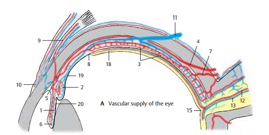
Vascular Supply
The eye has two different vascular systems: the ciliary arteries and the central retinalartery. All these vessels arise from theoph-thalmic artery. The posteriorciliary arteries are the branches supplying the vascular tunic of the eyeball, or uvea, which forms the iris (A1), the ciliary body (A2), and the choroidea proper of the poste-rior wall of the eyeball (A3). This vascular system not only supplies the blood, it is also essential for maintaining the intraocular pressure and the tension of the eyeball.
Long posterior ciliary arteries (A4). Thesearteries penetrate the sclera near the exit of the optic nerve. One of them runs in the temporal, the other one in the nasal wall of the eyeball to the ciliary body and to the iris. At the root of the iris they form thegreaterarterial circle of the iris (A5), from wherevessels radiate to the lesser arterial circle ofthe iris (A6) near the pupil.
Short posterior ciliary arteries (A7). Theseform the vascular plexus of the choroidea, which extends from the posterior wall of the eyeball to the oraserrata (A8). The inner choroid layer consists of especially wide capillaries, the choriocapillary layer, and borders on the pigmented epithelium of the retina. Whereas the choriocapillary lamina is firmly attached to the pigmented epithelium, the outer aspect of the choroidea is separated from the sclera by the perichoroidal space and, thus, can be dis-placed.
Anterior ciliary arteries (A9). These runfrom the rectus muscles to the sclera where they branch in the episcleral tissue and in the conjunctiva. In the conjunctiva they form the marginal loops (A10) around the margin of the cornea.The veins unite to form the fourposterior cil-iary veins, orvorticose veins(A11), whichobliquely penetrate the sclera at the poste-rior wall of the eyeball.

Central retinal artery (A12). This artery en-ters the optic nerve approximately 1 cm be-hind the eyeball and extends in the middleof the nerve to the papilla of the optic nerve (see below). It then divides into branches which run along the inner surface of the ret-ina within the layer of nerve fibers. The reti-nal vessels are end arteries. Their capillaries reach as far as the inner nuclear layer (see p. 349, A12). The venules unite to form the central vein of the retina (A13), which takes acourse similar to that of the central artery.
The visual cells are nourished from both sides of the retina: from the outside by the capillary system of the short posterior cili-ary arteries, and from the inside by the cen-tral arteries.
A18 Optical part of the retina.
A19 Ciliary part of the retina.
A20 Iridial part of the retina.
Related Topics