Home | | Human Nervous System and Sensory Organs | Orbital cavity (eye socket) - Structure of the Eye
Chapter: Human Nervous System and Sensory Organs : The Eye
Orbital cavity (eye socket) - Structure of the Eye

The orbital cavity (eye socket) is lined by the periosteum (periorbita) (C26) and is filled with fatty tissue.
Orbit
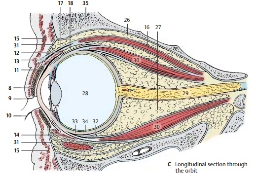
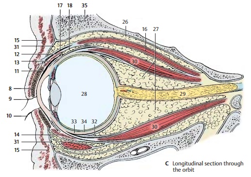
The orbital cavity (eye socket) is lined by the periosteum (periorbita) (C26) and is filled with fatty tissue, the orbital fat body (C27), in which the eyeball (C28), the optic nerve (C29), and the eye muscles (C30) are embedded. At the anterior margin of the orbit, the fatty tissue is demarcated by the orbital septum (BC31). The fatty tissue is separated from the eyeball by a connective tissue capsule, the bulbar sheath (C32), which encloses the sclera (C33).
C34 Choroidea.
C35 Osseous wall of the orbit.

Study Material, Lecturing Notes, Assignment, Reference, Wiki description explanation, brief detail
Human Nervous System and Sensory Organs : The Eye : Orbital cavity (eye socket) - Structure of the Eye |
Related Topics
Human Nervous System and Sensory Organs : The Eye