Chapter: Clinical Dermatology: Skin tumours
Tumours of the dermis: Malformations
Malformations
‘Salmon’ patches (‘stork bites’)
These
common malformations, present in about 50% of all babies, are caused by
dilatated capillaries in the superficial dermis. They are dull red, often
telangiec-tatic macules, most commonly on the nape of the neck (‘erythema
nuchae’), the forehead and the upper eyelids. Nuchal lesions may remain
unchanged, but patches in other areas usually disappear within a year.
‘Port-wine’ stains
These
are also present at birth and are caused by dilatated dermal capillaries. They
are pale, pink to purple macules, and vary from the barely noticeable to the
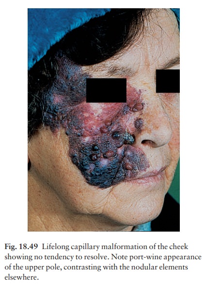
grossly disfiguring. Most occur on the face or trunk. They persist, and in
middle age may darken and become studded with angiomatous nodules (Fig. 18.49).
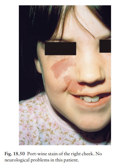
Occasionally a port-wine stain of the trigeminal area (Fig. 18.50) is
associated with a vascu-lar malformation of the leptomeninges on the same side,
which may cause epilepsy or hemiparesis (the Sturge–Weber syndrome), or with
glaucoma.

Excellent
results have been obtained with careful aand time-consumingatreatment with a
585-nm flashlamp-pumped pulsed dye laser. Treat-ment sessions can begin in
babies and anaesthesia is not always necessary. If a trial patch is
satisfactory, 40–50 pulses can be delivered in a session and the procedure can
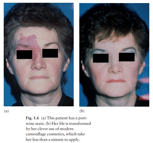
be repeated at 3-monthly intervals. On the other hand, some adults become very
adept at using cosmetic camouflage (see Fig. 1.6).

Combined vascular malformations of the limbs
A
large port-wine stain of a limb may be associated with overgrowth of all the
soft tissues of that limb with or without bony hypertrophy. There may be
underlying venous malformations (Klippel–Trenaunay syndrome), arteriovenous
fistulae (Parkes Weber syn-drome) or mixed venous–lymphatic malformations.

Related Topics