Chapter: Essentials of Anatomy and Physiology: Muscular System
Trunk Muscles
Trunk Muscles
Trunk muscles include those that move the vertebral column, the thorax and abdominal wall, and the pelvic floor.
Muscles Moving the Vertebral Column
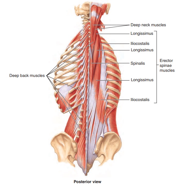
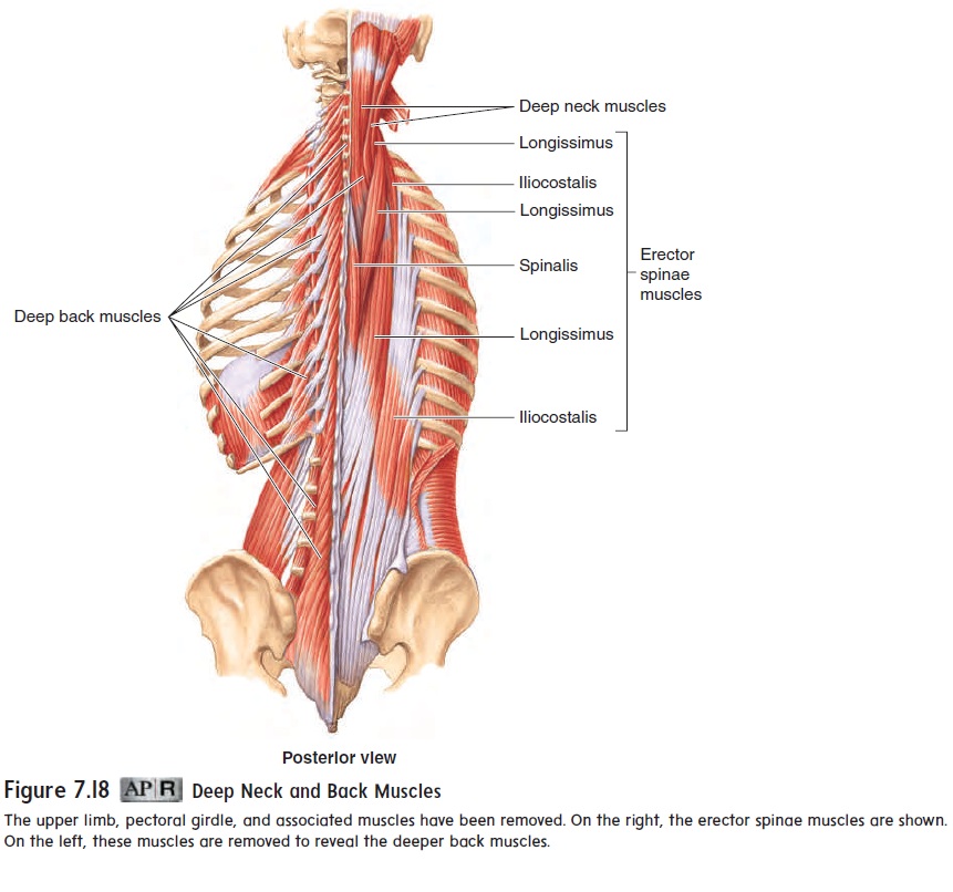
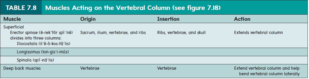
In humans, the back muscles are very strong to maintain erect posture. The erector spinae (ē-rek′tōr spı̄′nē) group of muscles on each side of the back are primarily responsible for keeping the back straight and the body erect (table 7.8 and figure 7.18). Deep back muscles, located between the spinous and transverseprocesses of adjacent vertebrae, are responsible for several movements of the vertebral column, including extension, lateral flexion, and rotation. When the deep back muscles are stretched abnormally or torn, muscle strains and sprains of lumbar verte-bral ligaments can occur, resulting in low back pain. Treatments include anti-inflammatory medication and RICE (rest, ice, compression, and elevation). Low back exercises can also helpthe problem.
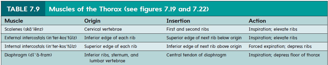
Thoracic Muscles
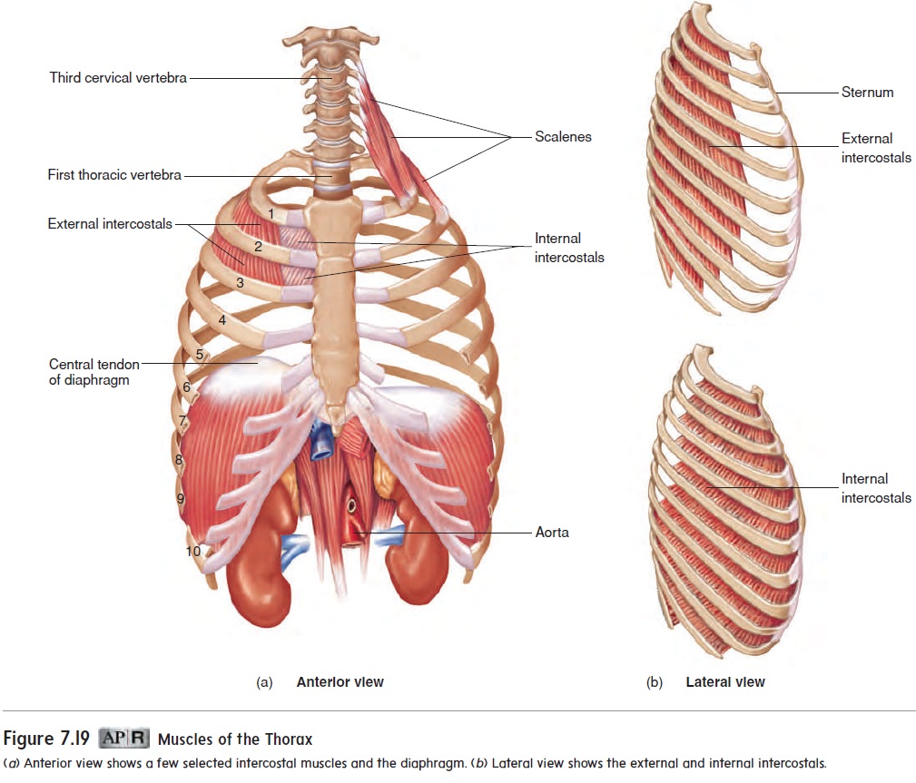
The muscles of the thorax (figure 7.19 and table 7.9) are involved almost entirely in the process of breathing. The external inter-costals (in′ter-kos′tŭlz; between ribs) elevate the ribs duringinspiration. Theinternal intercostals contract during forced expiration, depressing the ribs. However, the major movement produced in the thorax during quiet breathing is accomplished by the dome-shaped diaphragm (dı̄′ă-fram). When it contracts, the dome is flattened, causing the volume of the thoracic cavity to increase, resulting in inspiration.
Abdominal Wall Muscles
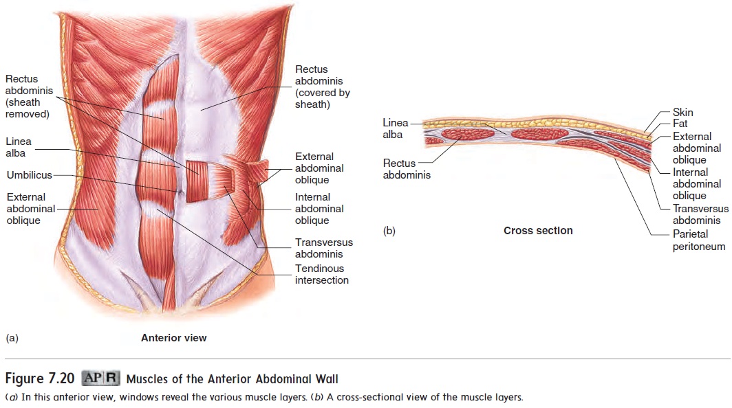
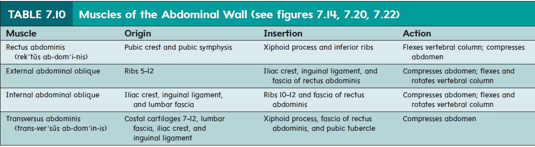
The muscles of the anterior abdominal wall (figure 7.20 and table 7.10) flex and rotate the vertebral column, compress the abdominal cavity, and hold in and protect the abdominal organs. In a relatively muscular person with little fat, a vertical indenta-tion, extending from the sternum through the navel to the pubis, is visible. This tendinous area of the abdominal wall, called the linea alba (lin′ē-ăal′bă; white line), consists of white connec-tive tissue rather than muscle. On each side of the linea alba is the rectus abdominis (rek′tŭs ab-dom′ ı̆-nis; rectus, straight) muscle. Tendinous intersections cross the rectus abdominis at three or more locations, causing the abdominal wall of a lean, well-muscled person to appear segmented. Lateral to the rectus abdominis are three layers of muscle. From superficial to deep, these muscles are the external abdominal oblique, the internalabdominal oblique, and the transversus abdominis (trans-ver′sŭs ab-dom′in-is) muscles. The fasciculi of these three muscle layers are oriented in different directions. When these muscles contract, they flex and rotate the vertebral column or compress the abdominal contents.
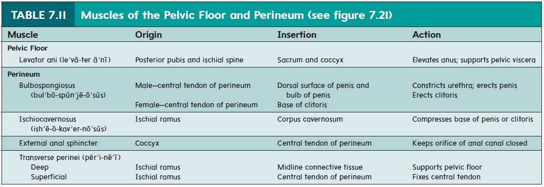
Pelvic Floor and Perineal Muscles
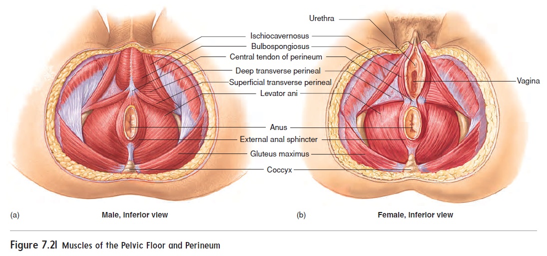
The pelvis is a ring of bone with an inferior opening that is closed by a muscular floor through which the anus and the openings of the urinary tract and reproductive tract penetrate. Most of the pelvicfloor, also referred to as thepelvic diaphragm,is formed by thelevator ani (le-vā′terā′nı̄) muscle. The area inferior to the pelvicfloor is the perineum (per′i-nē′ŭm), which contains a number of muscles associated with the male or female reproductive struc-tures (figure 7.21 and table 7.11). Several of these muscles help regulate urination and defecation.
Related Topics