Chapter: Essentials of Anatomy and Physiology: Muscular System
Excitability of Muscle Fibers
Excitability of Muscle Fibers
Muscle fibers, like other cells of the body, have electrical properties. This section describes the electrical properties of skeletal muscle fibers, and later sections illustrate their role in contraction.
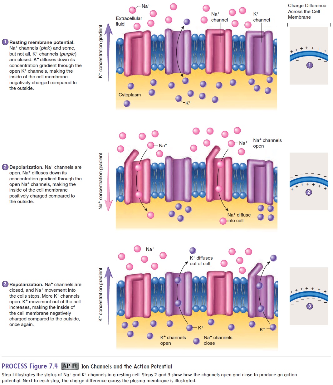
Most cells in the body have an electrical charge difference across their cell membranes. The inside of the membrane is negatively charged while the outside of the cell membrane is positively charged. In other words, the cell membrane is polar-ized (figure 7.4, step 1). The charge difference, called the resting membrane potential, occurs because there is an uneven distri-bution of ions across the cell membrane. The resting membrane potential develops for three reasons: (1) The concentration of K+ inside the cell membrane is higher than that outside the cell membrane; (2) the concentration of Na+ outside the cell mem-brane is higher than that inside the cell membrane; and (3) the cell membrane is more permeable to K+ than it is to Na+. Recall the different types of ion channels: nongated, or leak, channels, which are always open, and chemically gated channels, which are closed until a chemical, such as a neurotrans-mitter, binds to them and stimulates them to open (see figure 3.5). Because excitable cells have many K+ leak channels, K+ leaks out of the cell faster than Na+ leaks into the cell. In other words, some K+ channels are open, whereas other ion channels, such as those for Na+, are closed. In addition, negatively charged molecules, such as proteins, are in essence “trapped” inside the cell because the cell membrane is impermeable to them. For these reasons, the inside of the cell membrane is more negatively charged than the outside of the cell membrane.

In addition to an outward concentration gradient for K+, there exists an inward electrical gradient for K+. The resting membrane potential results from the equilibrium of K+ movement across the cell membrane. Because K+ is positively charged, its movement from inside the cell to the outside causes the inside of the cell membrane to become even more negatively charged compared to the outside of the cell membrane. However, potassium diffuses down its concentration gradient only until the charge difference across the cell membrane is great enough to prevent any additional diffusion of K+ out of the cell. The resting membrane potential is an equilibrium in which the tendency for K+ to diffuse out of the cell is opposed by the negative charges inside the cell, which tend to attract the positively charged K+ into the cell. At rest, the sodium-potassium pump transports K+ from outside the cell to the inside and transports Na+ from inside the cell to the outside. The active transport of Na+ and K+ by the sodium-potassium pump maintains the uneven distribution of Na+ and K+ across the cell membrane .
A change in resting membrane potential is achieved by changes in membrane permeability to Na+ or K+ ions. A stimula-tion in a muscle fiber or nerve cell causes Na+ channels to open
Because the Na+ concentration is much greater outside the cell than inside and the charge inside the cell membrane is negative, some positively charged Na+ quickly diffuses down its concentration gradient and toward the negative charges inside the cell, causing the inside of the cell membrane to become more positive than the outside of the cell. This change in the membrane potential is called depolarization. Near the end of depolarization, Na+ channels close, and additional K + channels open (figure 7.4, step 3). Consequently, the tendency for Na+ to enter the cell decreases, and the tendency for K+ to leave the cell increases. These changes cause the inside of the cell membrane to become more negative than the outside once again. Additional K+ channels then close as the charge across the cell membrane returns to its resting condition (figure 7.4, step 1). The change back to the resting membrane potential is called repolarization. The rapid depolarization and repolarization of the cell membrane is called an action potential. In a muscle fiber, an action potential results in muscle contraction.
Related Topics