Chapter: Essentials of Anatomy and Physiology: Tissues
Tissue membranes
Tissue membranes
List the structural and functional characteristics of mucous,serous, and synovial membranes.
A membrane is a thin sheet or layer of tissue that covers a struc-ture or lines a cavity. Most membranes consist of epithelium and the connective tissue on which the epithelium rests. There are four tissue membranes in the body. The skin, or cutaneous (kū-tā′\nē-ŭs, skin) membrane, is the external membrane\. It is composed of stratified squamous epithelium and dense connective tissue . The three major categories of internal membranes are mucous, serous, and synovial membranes.
Mucous Membranes
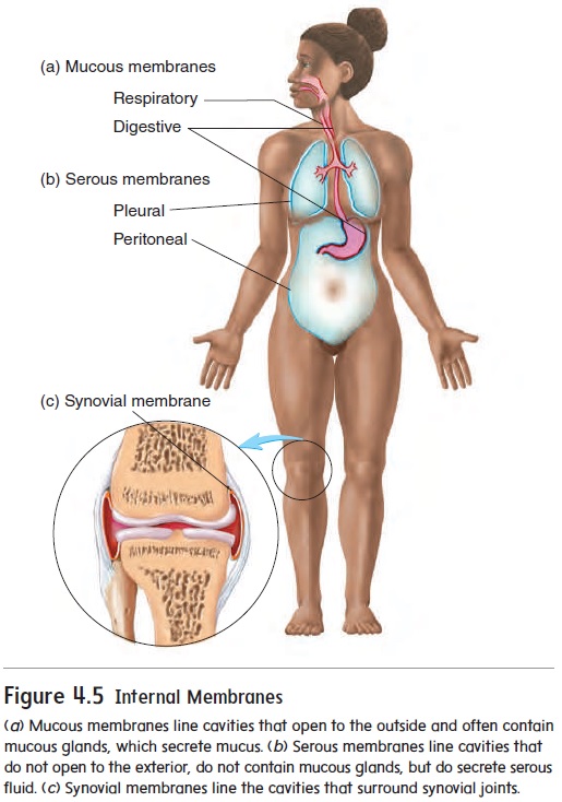
Mucous (mū′\kŭs) membranes consist of various kinds of epithe-lium resting on a thick layer of loose connective tissue. They line cavities that open to the outside of the body, such as the diges-tive, respiratory, and reproductive tracts (figure 4.5a). Many, but not all, mucous membranes have mucous glands, which secrete mucus.
The functions of mucous membranes vary, depending on their location, but they include protection, absorption, and secretion. For example, the stratified squamous epithelium of the oral cavity (mouth) performs a protective function, whereas the simple columnar epithelium of the intestine absorbs nutrients and secretes digestive enzymes and mucus. Mucous membranes also line the nasal passages. When it becomes inflamed, we experience the “runny nose” characteristic of the common cold or an allergy.
Serous Membranes
Serous (sēr′\ŭs; producing watery secretion) membranes consist ofsimple squamous epithelium resting on a delicate layer of loose con-nective tissue. Serous membranes line the trunk cavities and cover the organs within these cavities (figure 4.5b). Serous membranes secrete serous fluid, which covers the surface of the membranes. The smooth surface of the epithelial cells of the serous membranes combined with the lubricating qualities of the serous fluid prevent damage from abrasion when organs in the thoracic or abdomi-nopelvic cavities rub against one another. Serous membranes are named according to their location: The pleural (ploor′\ăl; a rib or the side) membranes are associated with the lungs; the pericardial (per-i-kar′\dē-ăl; around the heart) membranes areassociated with the heart; and the peritoneal (per′\i-tō-nē′\ăl; to stretch over) membranes are located in the abdominopelvic cavity (see figure 1.15). When the suffix -itis is added to the name of a structure, it means that the structure is inflamed (however, not all cases use the -itis suffix). Thus, pericarditis (per′\i-kar-dı̄′\tis) and peritonitis (per′\i-tō-n ı̄′\tis) refer to inflammation of the pericardial\ membranes and peritoneal membranes, respectively. Pleurisy (ploor′\i-sē) is inflammation of the pleural membranes.
(a) Mucous membranes line cavities that open to the outside and often contain mucous glands, which secrete mucus. (b) Serous membranes line cavities that do not open to the exterior, do not contain mucous glands, but do secrete serous fluid. (c) Synovial membranes line the cavities that surround synovial joints.
Synovial Membranes
Synovial (si-nō′\vē-ăl) membranes are made up of only con-nective tissue. They line the inside of joint cavities (the space where bones come together within a movable joint) (figure 4.5c). Synovial membranes produce synovial fluid, which makes the joint very slippery, thereby reducing friction and allowing smooth movement within the joint.
Related Topics