Chapter: Essentials of Anatomy and Physiology: Tissues
Connective tissue
Connective tissue
Describe the classification of connective tissue, and giveexamples of each major type.
Connective tissue is found throughout the body. It is usually char-acterized by large amounts of extracellular material that separates cells from one another. The extracellular material, or extracellularmatrix(mā′\triks), has three major components: (1) protein fibers,(2) ground substance consisting of nonfibrous protein and other molecules, and (3) fluid.
Three types of protein fibers help form most connective tissues. Collagen (kol′\lă-jen; glue-producing) fibers, which resemblemicroscopic ropes, are flexible but resist stretching. Reticular (rē-tik′\ū-lăr) fibersare very fine, short collagen fibers that branch to form a supporting network. Elastic fibers have a structure similar to that of coiled metal bed springs; after being stretched, they can recoil to their original shape.
Ground substance is the shapeless background against which cells and collagen fibers can be seen when using a light microscope. Although ground substance appears shapeless, the molecules within it are highly structured. Proteoglycans (prō′\tē-ō-glı̄′\kanz; proteo, protein + glycan, polysaccharide) resemble the limbs of pine trees, with proteins forming the branches and polysaccharides forming the pine needles. This structure enables proteoglycans to trap large quantities of water between the polysaccharides.
Connective tissue cells are named according to their functions. Cells whose names contain the suffix -blast (germ) produce the matrix; cells ending in -cyte (cell) maintain it; and cells ending in -clast (break) break it down for remodeling. For example, fibro-blasts (fı̄′\bro-blasts;fibra,fiber) are cells that form fibers andground substance in the extracellular matrix of fibrous connec-tive tissue, and fibroyctes are cells that maintain it. Osteoblasts (os′\tē-ō-blasts; osteo, bone) form bone, osteocytes (os′\tē-ō-sı̄tz) maintain bone, and osteoclasts (os′\tē-ō-klasts) break down bone.
\ Also found in connective tissue are cells associated with the immune system. Macrophages (mak′\rō-fāj-ez; makros, large + phago, to eat) are large white blood cells that are capable of movingabout and ingesting foreign substances, including microorganisms, in the connective tissue. Mast cells are nonmotile cells that release chemicals, such as histamine, that promote inflammation.
Functions of Connective Tissue
Connective tissue performs the following major functions:
1.Enclosing and separating other tissues. Sheets of connective tissue form capsules around organs, such as the liver and the kidneys. Connective tissue also forms layers that separate tissues and organs. For example, connective tissues separate muscles, arteries, veins, and nerves from one another.
2.Connecting tissues to one another. Tendons are strong cables, or bands, of connective tissue that attach muscles to bone, and ligaments are connective tissue bands that hold bones together.
3.Supporting and moving parts of the body. Bones of the skeletal system provide rigid support for the body, and semirigid cartilage supports structures, such as the nose, the ears, and the surfaces of joints. Joints between bones allow one part of the body to move relative to other parts.
4.Storing compounds. Adipose tissue (fat) stores high-energy molecules, and bones store minerals, such as calcium and phosphate.
5.Cushioning and insulating. Adipose tissue cushions and protects the tissues it surrounds and provides an insulating layer beneath the skin that helps conserve heat.
6.Transporting. Blood transports gases, nutrients, enzymes, hormones, and cells of the immune system throughout the body.
7Protecting. Cells of the immune system and blood provide protection against toxins and tissue injury, as well as against microorganisms. Bones protect underlying structures from injury.
Classification of Connective Tissue
Connective tissue types blend into one another, and the transition points cannot be precisely identified. As a result, connective tissue is somewhat arbitrarily classified by the type and proportions of cells and extracellular matrix.
Two major categories of connective tissue are embryonic and adult connective tissue. By eight weeks of development, most of the embryonic connective tissue has become specialized to form the types of connective tissue seen in adults.
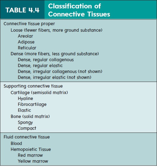
Table 4.4 presents the classification of adult connective tissue used in this text. Adult connective tissue consists of three types: connective tissue proper (loose and dense), supporting connective tissue (cartilage and bone), and fluid connective tissue (blood).
Connective Tissue Proper
Loose Connective Tissue
Loose connective tissue (table 4.5) consists of relatively fewprotein fibers that form a lacy network, with numerous spaces filled with ground substance and fluid. Three subdivisions of loose connective tissue are areolar, adipose, and reticular. Areolar (a-re′\ō-lar) has extracellular matrix consisting mostly of collagen fibers and a few elastic fibers. The most common cells in loose connective tissue are the fibroblasts, which are responsible for producing the matrix. Loose connective tissue is widely distributed
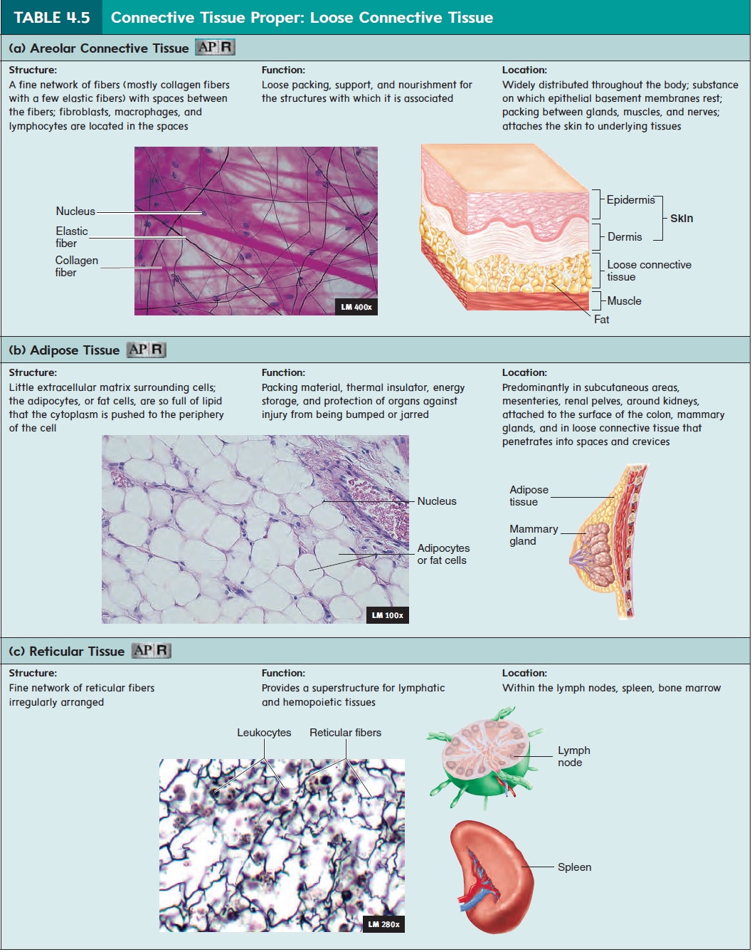
throughout the body and is the “loose packing” material of most organs and other tissues; it attaches the skin to underlying tissues and provides nourishment for the structures with which it is asso-ciated (table 4.5a). The basement membranes of epithelia often rest on loose connective tissue.
Adipose (ad′\i-pōs; fat) tissue consists of adipocytes, or fat cells, which contain large amounts of lipid for energy storage. Unlike other connective tissue types, adipose tissue is composed of large cells and a small amount of extracellular matrix, which consists of loosely arranged collagen and reticular fibers with some scattered elastic fibers. The individual cells are large and closely packed together (table 4.5b). Adipose tissue also pads andprotects parts of the body and acts as a thermal insulator.
Reticular tissue forms the framework of lymphatic tissue,such as in the spleen and lymph nodes, as well as in bone marrow and the liver (table 4.5c).
Dense Connective Tissue
Dense connective tissue has a relatively large number of protein fibersthat form thick bundles and fill nearly all of the extracellular space. These protein fibers are produced by fibroblasts. There are two major subcategories of dense connective tissue: collagenous and elastic. \
Dense collagenous connective tissue has an extracellular matrix consisting mostly of collagen fibers (table 4.6a). Structures made up of dense collagenous connective tissue include tendons, which attach muscle to bone; many ligaments, which attach bones to other bones; and much of the dermis, which is the connective tissue of the skin. Dense collagenous connective tissue also forms many capsules that surround organs, such as the liver and kidneys. In tendons and ligaments, the collagen fibers are oriented in the same direction, and so the tissue is called dense regular, but in the dermis and in organ capsules, the fibers are oriented in many different directions, and so the tissue is called dense irregular.
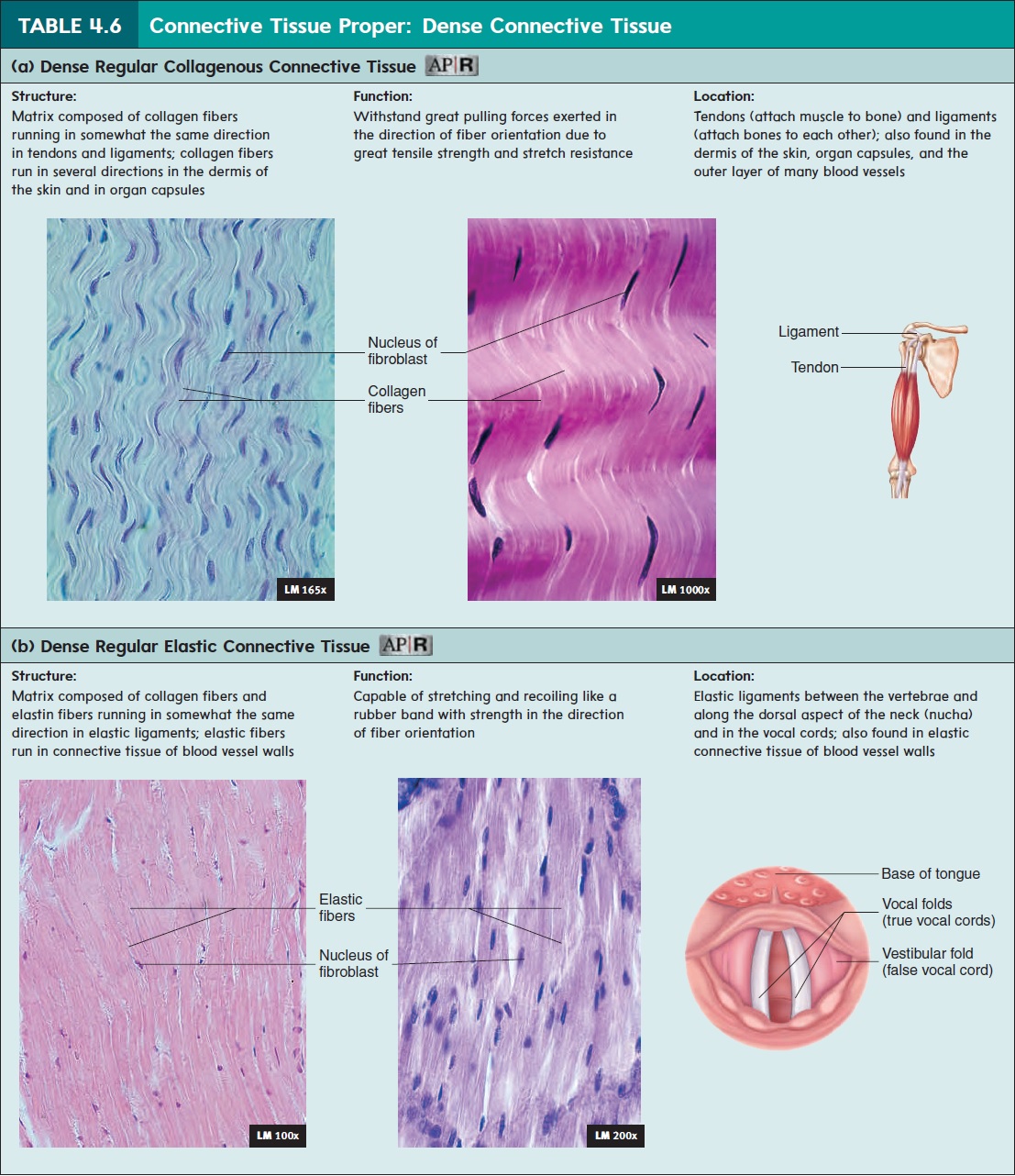
Dense elastic connective tissue has abundant elastic fibersamong its collagen fibers. The elastic fibers allow the tissue to stretch and recoil. Examples include the dense elastic connective tissue in the vocal cords (table 4.6b), in elastic ligaments, and in the walls of large arteries. The elastic fibers are oriented in the same direction in elastic ligaments and in the vocal cords (dense regular tissue), but they are oriented in many different directions in the walls of arteries (dense irregular tissue; not pictured).
Supporting Connective Tissue
Cartilage
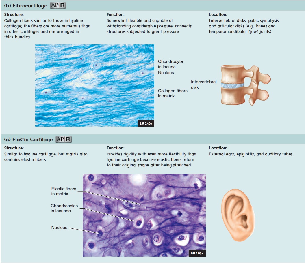
Cartilage (kar′ti-lij) is composed of chondrocytes (kon′dr̄o-s̄ıtz),or cartilage cells, located in spaces called lacunae (l̆a -koo′ n̄e ; small spaces) within an extensive matrix (table 4.7). Collagen in the matrix gives cartilage flexibility and strength. Cartilage is resilient because the proteoglycans of the matrix trap water, which makes the cartilage relatively rigid and enables it to spring back after being compressed. Cartilage provides support, but if bent or slightly compressed, it resumes its original shape. Cartilage heals slowly after an injury because blood vessels do not penetrate it. Thus, the cells and nutri-ents necessary for tissue repair do not easily reach the damaged area.
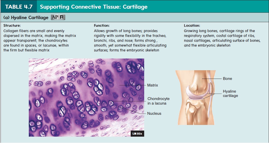
There are three types of cartilage:
1. Hyaline (h̄ı′̆a-lin; clear or glassy) cartilage (see table 4.7a)is the most abundant type of cartilage and has many functions. It covers the ends of bones where they come together to form joints. In joints, hyaline cartilage forms smooth, resilient surfaces that can withstand repeated compression. Hyaline cartilage also forms the cartilage rings of the respiratory tract, the nasal cartilages, and the costal cartilages, which attach the ribs to the sternum (breastbone).
3. Elastic cartilage (table 4.7c) contains elastic fibers inaddition to collagen and proteoglycans. The elastic fibers appear as coiled fibers among bundles of collagen fibers. Elastic cartilage is able to recoil to its original shape when bent. The external ear, epiglottis, and auditory tube contain elastic cartilage.
Bone
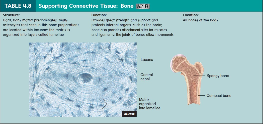
Bone is a hard connective tissue that consists of living cells and amineralized matrix (table 4.8). Osteocytes (osteo, bone), or bone cells, are located within lacunae. The strength and rigidity of the mineralized matrix enables bones to support and protect other tissues and organs. The two types of bone are compact bone and spongy bone
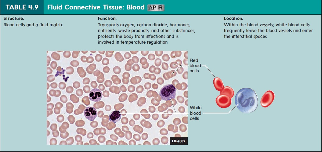
Blood
Blood is unique because the matrix is liquid, enabling blood cellsto move through blood vessels (table 4.9). Some blood cells even leave the blood and wander into other tissues. The liquid matrix enables blood to flow rapidly through the body, carrying nutrients, oxygen, waste products, and other materials.
Related Topics