Chapter: Essentials of Anatomy and Physiology: Tissues
Epithelial Tissue
EPITHELIAL TISSUE
1. List and explain the general characteristics of epithelial tissue.
2. Classify epithelial tissues based on the number of cell layers and the shape of the cells.
3. Name and describe the various types of epithelial tissue,including their chief functions and locations.
4. Relate the structural specializations of epithelial tissue withthe functions they perform.
5. Differentiate between exocrine and endocrine glands, andunicellular and multicellular glands.
6. Categorize glands based on their structure and function.
Epithelium (ep-i-thē′\lē-ŭm; pl. epithelia, ep-i-thē′\lē-ă;epi,on+ thele, covering or lining), or epithelial tissue, covers external and internal surfaces throughout the body. Surfaces of the body include the outer layer of the skin and the lining of cavities, such as the digestive tract, airways, and blood vessels. It also forms most glands. Epithelium consists almost entirely of cells with very little extracellular material between them. Although there are some exceptions, most epithelia have a free surface, which is not in contact with other cells, and a basal surface adjacent to a base-ment membrane, which attaches the epithelial cells to underlying tissues (figure 4.1). Epithelium may consist of a single layer of epithelial cells or multiple layers of epithelial cells between the free surface and the basement membrane.
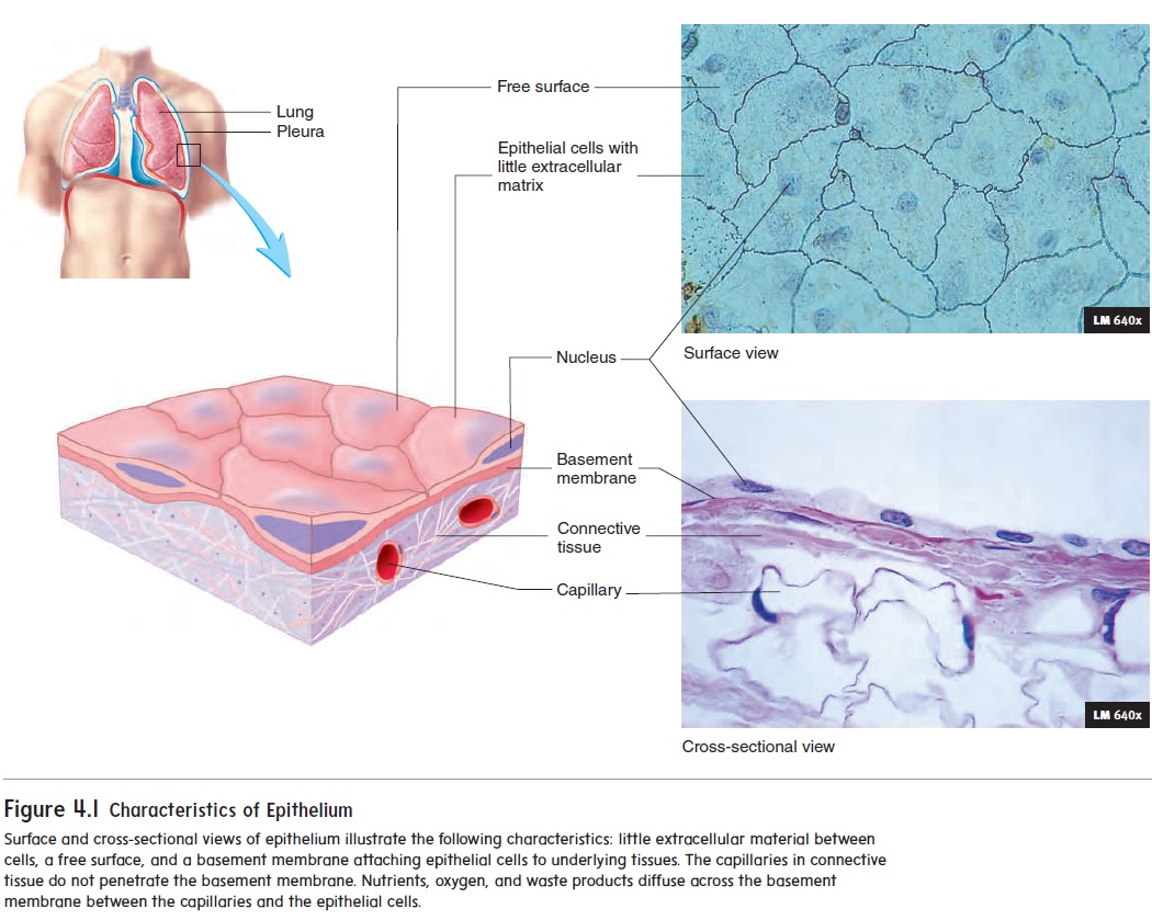
Figure 4.1 Characteristics of Epithelium
The basement membrane is secreted partly by epithelial cells and partly by the cells of the underlying tissues. It consists of a meshwork of protein molecules with other molecules bound to them. Substances that cross the epithelium must also cross the basement membrane. It can function as a filter and as a barrier to the movement of cells. For example, if some epithelial cells are converted to cancer cells, the basement membrane can, for some time, help prevent the spread of the cancer into the underlying tissues.
Blood vessels do not extend from the underlying tissues into epithelium, so gases and nutrients that reach the epithelium must diffuse across the basement membrane from the underlying tissues, where blood vessels are abundant. Waste products produced by the epithelial cells diffuse across the basement membrane in the opposite direction, to reach the blood vessels.
Functions of Epithelia
The major functions of epithelia are
1.Protecting underlying structures. Examples include the outer layer of the skin and the epithelium of the oral cavity, which protect the underlying structures from abrasion.
2.Acting as a barrier. Epithelium prevents many substances from moving through it. For example, the epithelium of the skin acts as a barrier to water and reduces water loss from the body. The epithelium of the skin also prevents many toxic molecules and microorganisms from entering the body.
3.Permitting the passage of substances. Epithelium also allows many substances to move through it. For example, oxygen and carbon dioxide are exchanged between the air and blood by diffusion through the epithelium in the lungs.
4.Secreting substances. Sweat glands, mucous glands, and the enzyme-secreting portion of the pancreas are all composed of epithelial cells.
5.Absorbing substances. The cell membranes of certain epithelial tissues contain carrier proteins that regulate the absorption of materials. For example, the epithelial cells of the intestines absorb digested food molecules, vitamins, and ions.
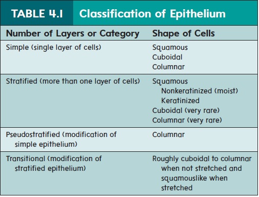
Classification of Epithelia
Epithelia are classified according to the number of cell layers and the shape of the cells (table 4.1). Simple epithelium consists of a single layer of cells. Stratified epithelium consists of more than one layer of epithelial cells, with some cells sitting on top of others. Categories of epithelium based on cell shape are squamous (skwā′\mŭs; flat), cuboidal (cubelike), and columnar (tall and thin). In most cases, each epithelium is given two names, such as simple squamous, simple columnar, or stratified squamous epithelium. When epithelium is stratified, it is named according to the shape of the cells at the free surface.
Simple squamous epithelium is a single layer of thin, flat cells (table 4.2a). Some substances easily pass through this thin layer of cells, but other substances do not. For example, the air-ways end as small sacs called alveoli (al-vē′\o-lı̄; sing. alveolus, hollow sac). The alveoli consist of simple squamous \epithelium that allows oxygen from the air to diffuse into the body and carbon dioxide to diffuse out of the body into the air. Simple squamous epithelium in the filtration membranes of the kidneys forms thin barriers through which small molecules, but not large ones, can pass. Small molecules, including water from blood, are filtered through these barriers as a major step in urine formation. Large molecules, such as proteins, and blood cells remain in the blood vessels of the kidneys.
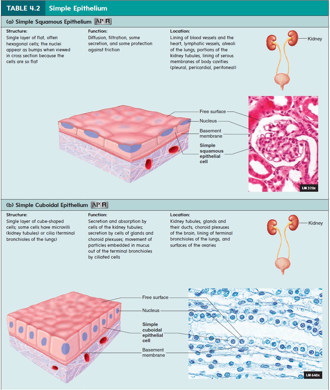
Simple squamous epithelium also prevents abrasion between organs in the pericardial, pleural, and peritoneal cavities . The outer surfaces of organs are covered with simple squamous epithelium that secretes a slippery fluid. The fluid lubri-cates the surfaces between the organs, preventing damage fromfriction when the organs rub against one another or the body wall.
Simple cuboidal epithelium is a single layer of cubelikecells (table 4.2b) that carry out active transport, facilitated dif-fusion, or secretion. Epithelial cells that secrete molecules such as proteins contain organelles that synthesize them. These cells have a greater volume than simple squamous epithelial cells and contain more cell organelles. The kidney tubules have large portions of their walls composed of simple cuboidal epithelium. These cells secrete waste products into the tubules and reabsorb useful materials from the tubules as urine is formed. Some cuboidal epithelial cells have cilia that move mucus over the free surface or microvilli that increase the surface area for secretion and absorption.
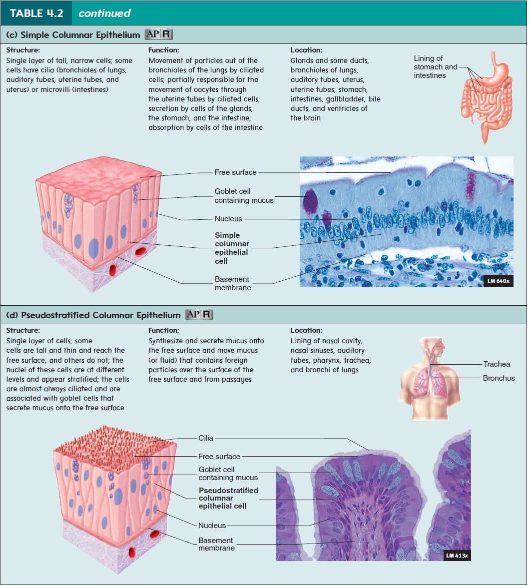
Pseudostratified columnar epithelium is a special type ofsimple epithelium (table 4.2d). The prefix pseudo- means false, so this type of epithelium appears stratified but is not. It consists of one layer of cells, with all the cells attached to the basement mem-brane. But it looks like two or more layers of cells because some of the cells are tall and reach the free surface, whereas others are short and do not reach the free surface. Pseudostratified columnar epithelium lines some glands and ducts, the auditory tubes, and some of the airways, such as the nasal cavity, nasal sinuses, phar-ynx, trachea, and bronchi. Pseudostratified columnar epithelium secretes mucus, which covers its free surface. Cilia located on the free surface move the mucus and the debris that accumulates in it. For example, cilia of the airways move mucus toward the throat, where it is swallowed.
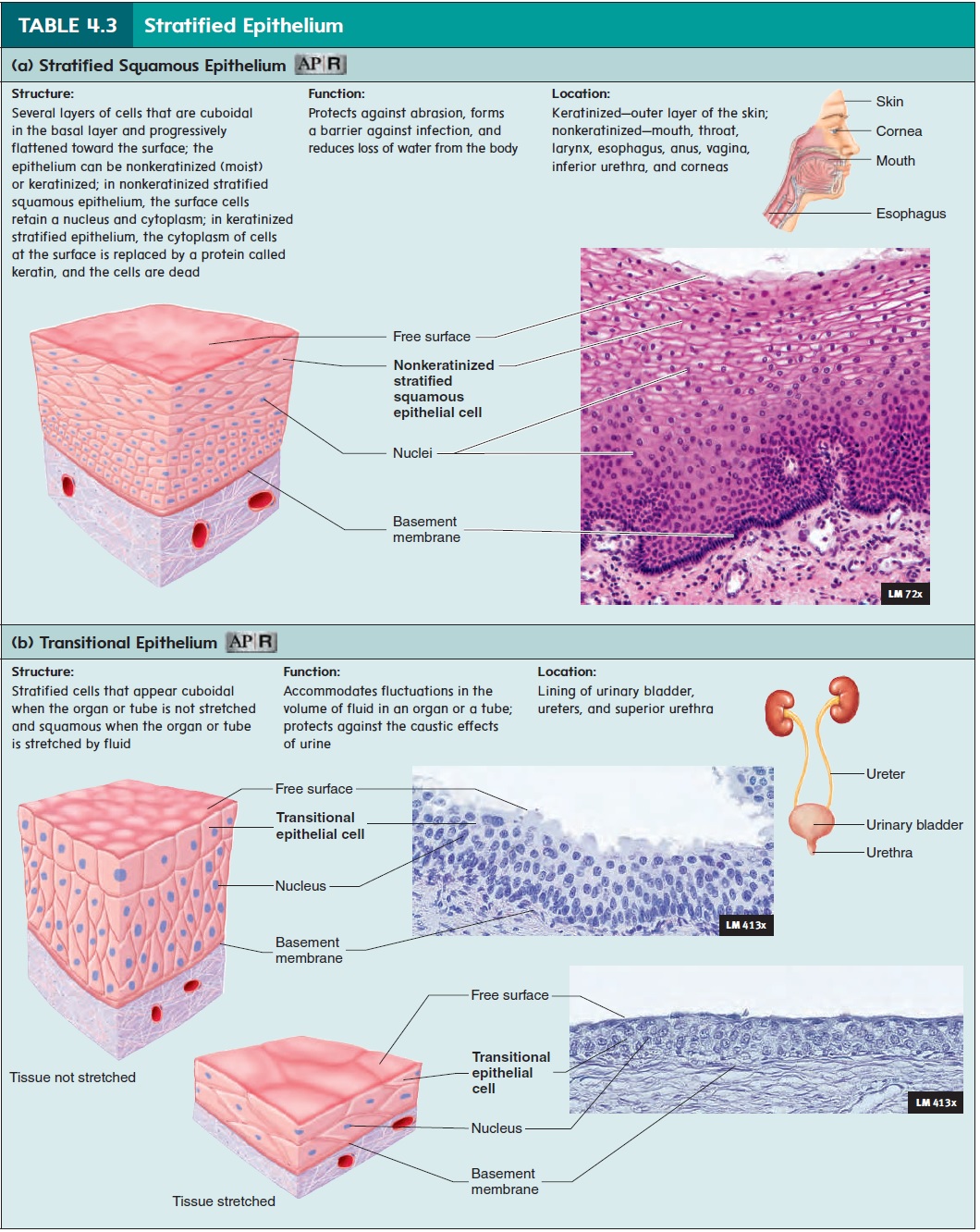
Stratified squamous epithelium forms a thick epithelium because it consists of several layers of cells (table 4.3a). The deepest cells are cuboidal or columnar and are capable of dividing and producing new cells. As these newly formed cells are pushed to the surface, they become flat and thin. As the cells flatten, the cyto-plasm of the epithelial cells is replaced by a protein called keratin, and the cells die. One type of stratified squamous epithelium forms the outer layer of the skin and is called keratinized squa-mous epithelium . The dead cells provide protection against abrasion, and form a barrier that prevents microorganisms and toxic chemicals from entering the body, and reduces the loss of water from the body. If cells at the surface are damaged or rubbed away, they are replaced by cells formed in the deeper layers.
In contrast, stratified squamous epithelium of the mouth is composed of living cells with a moist surface. This nonkeratinized (moist) stratified squamous epithelium also provides protection against abrasion and acts as a mechanical barrier, preventing microorganisms from entering the body. Water, however, canmove across it more readily than across the skin.
Stratified cuboidal epithelium consists of more than onelayer of cuboidal epithelial cells. This epithelial type is relatively rare and is found in sweat gland ducts, ovarian follicular cells, and the salivary glands. It functions in absorption, secretion, and protection.
Stratified columnar epithelium consists of more than one layer of epithelial cells, but only the surface cells are columnar. The deeper layers are irregular or cuboidal in shape. Like stratified cuboidal epithelium, stratified columnar epithelium is relatively rare. It is found in the mammary gland ducts, the larynx, and a portion of the male urethra. This epithelium carries out secretion,protection, and some absorption.
Transitional epithelium is a special type of stratified epithe-lium that can be greatly stretched (table 4.3b). In the unstretched state, transitional epithelium consists of five or more layers of cuboidal or columnar cells that often are dome-shaped at the free surface. As transitional epithelium is stretched, the cells change to a low cuboidal or squamous shape, and the number of cell layers decreases. Transitional epithelium lines cavities that can expand greatly, such as the urinary bladder. It also protects underlying structures from the caustic effects of urine.
Structural and Functional Relationships
Cell Layers and Cell Shapes
The number of cell layers and the shape of the cells in a specific type of epithelium reflect the function the epithelium performs. Two important functions are controlling the passage of materials through the epithelium and protecting the underlying tissues. Simple epithelium, with its single layer of cells, is found in organs that primarily function to move materials. Examples include the diffusion of gases across the wall of the alveoli of the lungs, filtra-tion of fluid across the filtration membranes in the kidneys, secretion from glands, and nutrient absorption by the intestines. The move-ment of materials through a stratified epithelium is hindered by its many layers. Stratified epithelium is well adapted for its protective function. As the outer cell layers are damaged, they are replaced by cells from deeper layers. Stratified squamous epithelium is found in areas of the body where abrasion can occur, such as in the skin, anal canal, and vagina.
Differences in function are also reflected in cell shape. Cells are normally flat and thin when the function is diffusion, such as in the alveoli of the lungs, or filtration, such as in kidney glomeruli. Cells with the major function of secretion or absorption are usu-ally cuboidal or columnar. They are larger because they contain more organelles, which are responsible for the function of the cell. The stomach, for example, is lined with simple columnar epithelium. These cells contain many secretory vesicles (ves′\i-klz) filled with mucus. The large amounts of mucus produced by the simple columnar epithelium protect the stomach lining against the digestive enzymes and acid produced in the stomach. An ulcer, or irritation in the stomach’s epithelium and underlying tissue, can develop if this protective mechanism fails. Simple cuboidal epithelial cells that secrete or absorb molecules, as occurs in the kidney tubules, contain many mitochondria, which produce the ATP required for active transport.
The shape and number of layers of epithelial cells can change if they are subjected to long-term irritation or other abnormal condi-tions. People who smoke cigarettes eventually experience changes in the epithelium of the larger airways. The delicate pseudostrati-fied columnar epithelium, which performs a cleaning function by moving mucus and debris from the \passageways, is replaced by stratified squamous epithelium, which is more resistant to irri-tation but does not perform a cleaning function. Also, lung cancer most often results from changes in epithelial cells in the lung passageways of smokers. The changes in cell structure are used to identify the cancer.
Free Surfaces
Most epithelia have a free surface that is not in contact with other cells and faces away from underlying tissues. The characteristics of the free surface reflect its functions. The free surface can be smooth or lined with microvilli or cilia. A smooth free surface reduces friction as material moves across it. For example, the lin-ing of blood vessels is simple squamous epithelium with a smooth surface, which reduces friction as blood flows through the vessels. Microvilli are cylindrical extensions of the cell membrane thatincrease the free surface area . Normally, many microvilli cover the free surface of each cell involved in absorp-tion or secretion, such as the cells lining the small intestine. Cilia propel materials along the free surface of cells. The nasal cavity and trachea are lined with pseudostratified columnar ciliated epithelium. Intermixed with the ciliated cells are special-ized mucus-producing cells called goblet cells (see table 4.2d). Dust and other materials are trapped in the mucus that covers the epithelium, and movement of the cilia propels the mucus with its entrapped particles to the back of the throat, where it is swallowed or coughed up. The constant movement of mucus helps keep the airways clean.
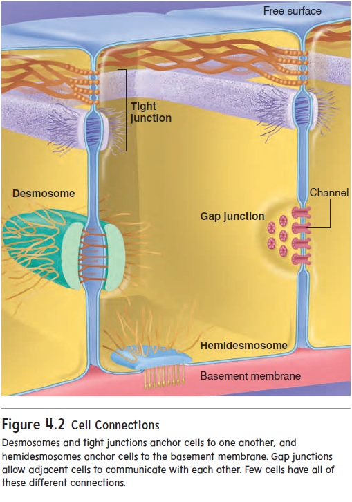
Cell Connections
Epithelial cells are connected to one another in several ways (figure 4.2). Tight junctions bind adjacent cells together and form permeability barriers. Tight junctions prevent the passage of materials\ between epithelial cells because they completely sur-round each cell, similar to the way a belt surrounds the waist. Materials that pass through the epithelial layer must pass through the cells, so those cells regulate what materials can cross. Tight junc-tions are found in the lining of the intestines and in most other simple epithelia. Desmosomes (dez′\mō-sōmz; desmos, a band + soma, body) are mechanical links that bind cells together. Modified des-mosomes, called hemidesmosomes (hem-ē-dez′\mō-sōmz; hemi, one-half), also anchor cells to the basement membrane. Many desmosomes are found in epithelia subjected to stress, such as the stratified squamous epithelium of the skin. Gap junctions are small channels that allow small molecules and ions to pass from one epithelial cell to an adjacent one. Most epithelial cells are connected to one another by gap junctions, and researchers believe that molecules or ions moving through the gap junctions act as communication signals to coordinate the activities of the cells.
Glands
A gland is a structure that secretes substances onto a surface, into a cavity, or into the blood. Most glands are composed primarily of epithelium and are multicellular. But sometimes single goblet cells are classified as unicellular glands because they secrete mucus onto epithelial surfaces.
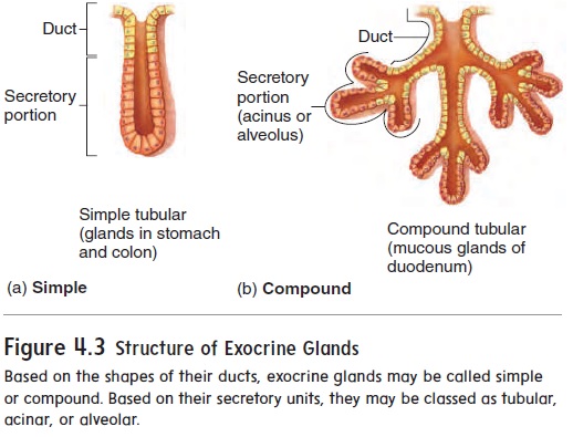
Glands with ducts are called exocrine (ek′\sō-krin; exo, outside + krino, to separate) glands (figure 4.3). The exocrine glands can be simple, with ducts that have no branches, or compound, with ducts that have many branches. The end of a duct can be tubular. Some tubular glands are straight, and others are coiled. Some glands have ends that are expanded into a saclike structure called an acinus (as′\i-nŭs; grapelike) or an alveolus (al-vē′\ō-lŭs; small cavity). Some compound glands have acini and tubules (tubuloacinar or tubuloalveolar glands) that secrete sub-stances. Secretions from exocrine glands pass through the ducts onto a surface or into an organ. For example, sweat from sweat glands and oil from sebaceous glands flow onto the skin surface.
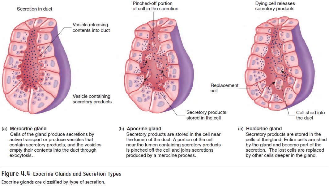
Exocrine glands can also be classified according to how products leave the cell. The most common type of secretion is merocrine (mer′\ōkrin) secretion. In merocrine secretion, productsare released, but no actual cellular material is lost (figure 4.4a). Secretions are either actively transported or packaged in vesicles and then released by the process of exocytosis at the free surface of the cell. Sweat and digestive enzymes produced by the pancreas are released by merocrine secretion. In apocrine (ap′\ō-krin) secretion, the secretory products are released as fragments of the gland cell (figure 4.4b). Milk secretion by the mammary glands utilizes some apocrine secretion. Holocrine (hol′\ō-krin) secretion involves the shedding of entire cells (figure 4.4c). Sebaceous (oil) glands of the skin utilize holocrine secretion.
Endocrine (en′\dō-krin; endo, within) glands have no ducts and empty their secretions into the blood. These secretions, called hormones (hōr′\mōnz), are carried by the blood to other partsof the body. Endocrine glands include the thyroid gland and the insulin-secreting portions of the pancreas.
Related Topics