Chapter: Essentials of Anatomy and Physiology: The Endocrine System
Thyroid Gland - Anatomy and Physiology
THYROID GLAND
The thyroid gland is located on the front and sides of the trachea just below the larynx. Its two lobes are connected by a middle piece called the isthmus. The structural units of the thyroid gland are thyroid folli-cles, which produce thyroxine (T4) and triiodothy-ronine (T3). Iodine is necessary for the synthesis of these hormones; thyroxine contains four atoms of iodine, and T3 contains three atoms of iodine.
The third hormone produced by the thyroid gland is calcitonin, which is secreted by parafollicular cells. Its function is very different from those of thyroxine and T3.
THYROXINE AND T3
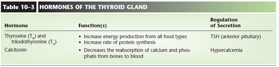
Thyroxine (T4) and T3 have the same functions: regulation of energy production and protein synthesis, which contribute to growth of the body and to normal body functioning throughout life (Fig. 10–5). Thyroxine and T3 increase cell respiration of all food types (carbohydrates, fats, and excess amino acids) and thereby increase energy and heat production. They also increase the rate of protein synthesis within cells. Normal production of thyroxine and T3 is essential for physical growth, normal mental development, and maturation of the reproductive system. These hormones are the most important day-to-day regulators of metabolic rate; their activity is reflected in the func
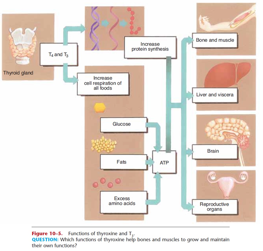
Figure 10–5. Functions of thyroxine and T3.
QUESTION: Which functions of thyroxine help bones and muscles to grow and maintain their own functions?
tioning of the brain, muscles, heart, and virtually all other organs. Although thyroxine and T3 are not vital hormones, in that they are not crucial to survival, their absence greatly diminishes physical and mental growth and abilities.
Secretion of thyroxine and T3 is stimulated by thyroid-stimulating hormone (TSH) from the ante-rior pituitary gland (see also Fig. 1–3). When the metabolic rate (energy production) decreases, this change is detected by the hypothalamus, which secretes thyrotropin releasing hormone (TRH). TRH stimulates the anterior pituitary to secrete TSH, which stimulates the thyroid to release thyroxine and T3, which raise the metabolic rate by increasing energy production. This negative feedback mecha-nism then shuts off TRH from the hypothalamus until the metabolic rate decreases again.
CALCITONIN
Calcitonin decreases the reabsorption of calcium and phosphate from the bones to the blood, thereby lowering blood levels of these minerals. This function of calcitonin helps maintain normal blood levels of calcium and phosphate and also helps maintain a stable, strong bone matrix. It is believed that calcitonin exerts its most important effects during childhood, when bones are growing. A form of calci-tonin obtained from salmon is used to help treat osteoporosis.
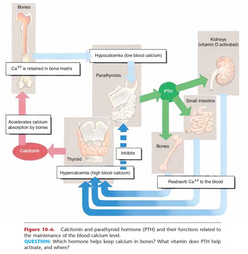
The stimulus for secretion of calcitonin is hyper-calcemia, that is, a high blood calcium level. When blood calcium is high, calcitonin ensures that no more calcium will be removed from bones until there is a real need for more calcium in the blood (Fig. 10–6). The hormones of the thyroid gland are summarized in Table 10–3.
Related Topics