Chapter: Basic Radiology : Imaging of the Heart and Great Vessels
Techniques and Normal Anatomy : Magnetic Resonance Imaging and Angiography
Magnetic
Resonance Imaging
MR imaging has also gained rapid
acceptance for cardiac eval-uation, as it does not use ionizing radiation, can
provide mor- phologic and physiologic data, and can be performed to give
cine-loop images. MR cardiac imaging remains a challenge be-cause of the
inherent difficulty of simultaneously dealing with respiratory and cardiac
motion, the competing needs for spa-tial and temporal data, and the hands-on
approach to tailor the examination to the specific clinical question. Thus, MR
imaging is largely a problem-solving tool, rather than a screening study. The
major indications for MR imaging are congenital heart disease and suspected
intracardiac masses, valvular dysfunc-tion, pericardial disease, and aortic
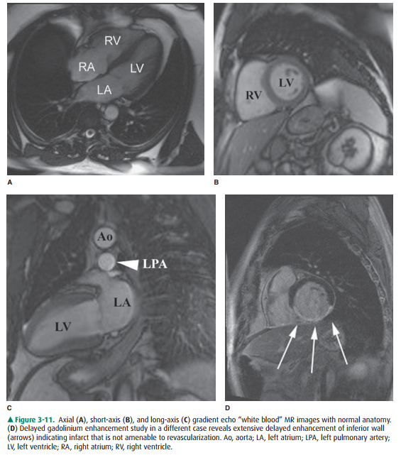
abnormality. From a func-tional standpoint, MR has the ability to asses cardiac
function and motion, distinguish infarct from ischemia and help deter-mine the
advisability of revascularization (Figure 3-11), and measure flow across valves
or coarctations. On the research side, MR imaging has also shown some promise
in measuring the degree of damage from coronary artery atherosclerosis and
evaluating the composition of atherosclerotic plaque.
Angiography
Conventional angiography is one
of the most commonly per-formed imaging tests for evaluating the heart and
great ves-sels. After the introduction of a catheter into a peripheral vessel
(usually, the femoral or axillary vein or artery), the an-giographer, under
fluoroscopic visualization, positions the catheter in the region of interest,
injects contrast material to confirm the location of the catheter, and then
injects larger amounts of contrast material for diagnostic purposes. This injection
of contrast material can be videotaped, recorded as standard or digital
radiographs, or digitally stored for later review. There are four major types
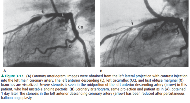
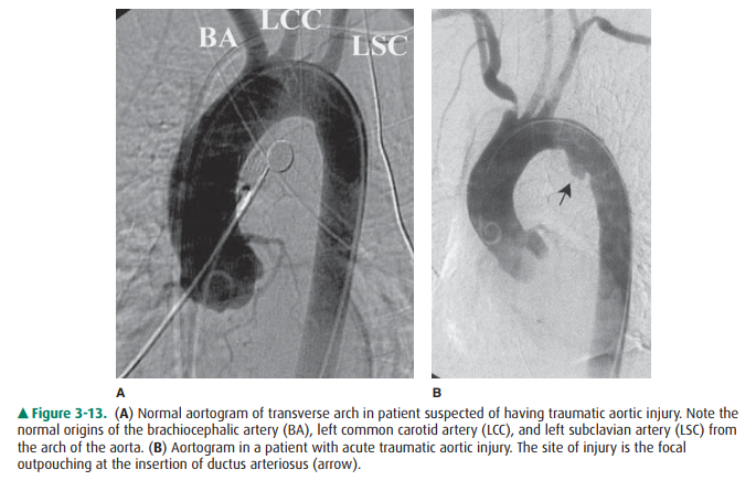
of angiography: angiocardiography (heart), coronary arteriography (coronary
arteries) (Figure 3-12), aortography (aorta) (Figure 3-13), and pulmonary
angiography (pulmonary arteries and lungs). Techniques developed by
radiologists, angiocardiography and coronary arteriography, are now almost
exclusively per-formed by cardiologists.
Related Topics