Chapter: Human Neuroanatomy(Fundamental and Clinical): Internal Structure of the Spinal Cord
Subdivisions of Neural Tube
SUBDIVISIONS OF NEURAL TUBE
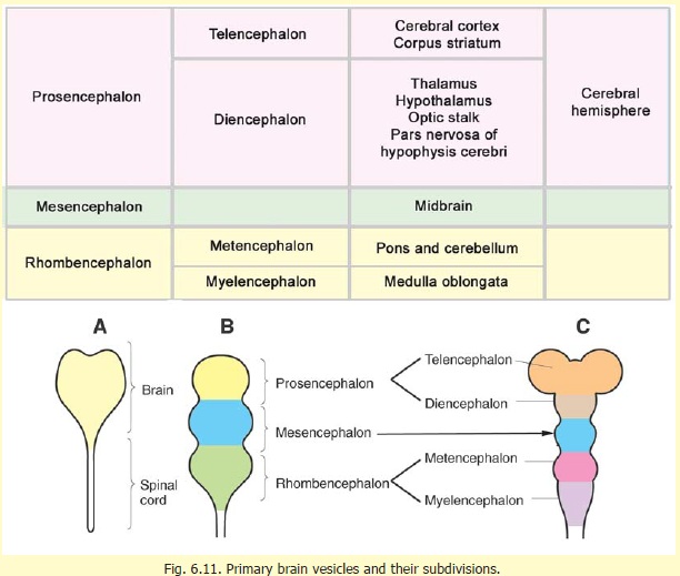
We have seen earlier that even before the neural tube has completely closed, it is divisible into an enlarged cranial part and a caudal tubular part. The enlarged cranial part forms the brain. The caudal tubular part forms the spinal cord: it is at first short, but gradually gains in length as the embryo grows. The cavity of the developing brain soon shows three dilatations (Fig. 6.11B). Cranio-caudally, these are the prosencephalon, mesencephalon, and rhombencephalon. The prosencephalon becomes subdivided into thetelencephalon and the diencephalon (Fig. 6.11C). The telencephalon consists of right and left telencephalic vesicles. The rhombencephalon also becomes subdivided into a cranial part, the metencephalon, and a caudal part, the myelencephalon. The parts of the brain that are developed from each of these divisions of the neural tube are shown in Fig. 6.11.
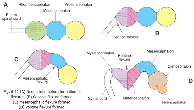
The prosencephalon, mesencephalon and rhombencephalon are at first arranged cranio-caudally (Fig. 6.12A). Their relative position is greatly altered by the appearance of a number of flexures. These are:
(a) the cervical flexure, at the junction of the rhombencephalon and the spinal cord (Fig. 6.12B);
(b) themesencephalic flexure(orcephalic flexure); in the region of the midbrain (Fig. 6.12C);
(c) the pontine flexure, at the middle of the rhombencephalon, dividing it into the metencephalon and myelencephalon (Fig. 6.12D); and
(d) thetelencephalic flexure, that occurs much later, between the telencephalon anddiencephalon.
These flexures lead to the orientation of the various parts of the brain as in the adult.
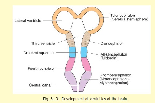
Each of the subdivisions of the developing brain encloses a part of the original cavity of the neural tube (Fig. 6.13). The cavity of each telencephalic vesicle becomes the lateral ventricle, and that of the diencephalon (along with the central part of the telencephalon), becomes the third ventricle. The cavity of the mesencephalon remains narrow, and forms the aqueduct, while the cavity of the rhombencephalon forms the fourth ventricle. Its continuation in the spinal cord is the central canal.
Related Topics