Chapter: Human Neuroanatomy(Fundamental and Clinical): Internal Structure of the Spinal Cord
Subdivisions of Grey Matter

Subdivisions of Grey Matter
The grey matter of the spinal cord may be subdivided in more than one manner.
Traditionally, the ventral grey column has been divided into a ventral part, the head, and a dorsal part, the base. Similarly, the dorsal grey column has been subdivided (from anterior to posterior side) into a base, a neck, and a head. These subdivisions have, however, been found to have little importance.
Nuclei in spinal grey matter
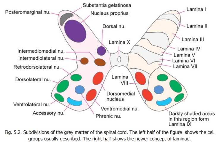
An attempt has been made to recognise discrete collections of neurons (or nuclei) in various regions of the spinal grey matter. These are illustrated in the left half of Fig. 5.2.
The neurons of the ventral grey column (ventral horn cells) are arranged in the form of several discrete and elongated groups. We can recognize
a. medial group which may be subdivided into dorsomedial and ventromedial parts;
b. lateral group consisting of dorsolateral, ventrolateral and retrodorsolateral parts;
c. acentral group represented by the phrenic and accessory nuclei (in the cervical region) and by the lumbosacral nucleus (in the lumbosacral region). Some large cells present along the anterior margin of the ventral grey column are called spinal border cells.
Within the dorsal grey column the following relatively discrete areas may be recognised.
i. The substantia gelatinosa which lies near the apex.
ii. Nucleus proprius (or dorsal funicular group).
iii. Dorsal nucleus (also called the thoracic nucleus or Clark’s column) lying on the medial side of the base.
iv. A thin layer of cells lying superficial to the substantia gelatinosa and constituting the posteromarginal nucleus, also called the marginal zone.
Between the ventral and dorsal grey columns an intermediate zone is sometimes described. This contains the intermediolateraland intermediomedial nuclei (or columns).
Advanced
In the right half of Fig. 5.2.the ventral grey column is shown to be subdivided by a line passing forwards and medially. The part of the column lateral to this line (containing the greater part of the lateral group of nuclei) is present only in the region of the cervical and lumbar enlargements of the spinal cord. From this it can be inferred that the lateral group of nuclei innervate the musculature of the limbs. (Some neurons occupying the position of the ventrolateral group are seen in the first and second sacral segments of the spinal cord. They are believed to supply perineal musculature).
The dorsal nucleus is present only in the thoracic and upper lumbar segments. The intermediolateral group of neurons is present (a) from segments T1 to L2, and (b) in segments S2, S3 and S4.
From the examples given above it will be clear that at a given level of the spinal cord only some of the nuclear groups named in the preceding paragraphs are present.

Division of spinal grey matter into laminae
Recent studies have shown that from the point of view of neuronal connections the grey matter of the spinal cord may be divided into ten areas or laminae. These are illustrated in the right half of Fig. 5.2.
Lamina I corresponds to the posteromarginal nucleus (lamina marginalis), lamina II to thesubstantia gelatinosa, and laminae IIIand IV to the nucleus proprius. (Laminae I to IV correspond to the head of the dorsal grey column. Some workers include lamina III in the substantia gelatinosa). Lamina V corresponds to the neck of the dorsal grey column and lamina VI to the base. The lateralpart of lamina V corresponds to the formatio reticularis. Lamina VII corresponds to the intermediate grey matter and includes the intermediomedial, intermediolateral, and dorsal nuclei. It is believed to be made up predominantly of interneurons. At the level of the cervical and lumbar enlargements this lamina extends into the lateral part of the ventral horn. Lamina VIII occupies most of the ventral horn in the thoracic segments, but at the level of the limb enlargements it is confined to the medial part of the ventral horn.Lamina IX is made up of the various discrete groups of ventral horn cells already described. Lamina X forms the grey matter around the central canal.
From the point of view of function, and of neuronal organisation, it has been proposed that (instead of division into ventral and dorsal grey columns) the spinal grey matter should be divided into a central core where the organisation of neurons is diffuse and non-discriminative; and into dorsal and ventral appendages. Laminae VII and VIII have been (tentatively) assigned to the centralcore, laminae I to VI to the dorsal appendage, and lamina IX to the ventral appendage.
Related Topics