Chapter: Human Neuroanatomy(Fundamental and Clinical): Internal Structure of the Spinal Cord
The Midbrain: gross anatomy
The Midbrain: gross anatomy
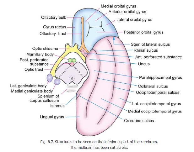
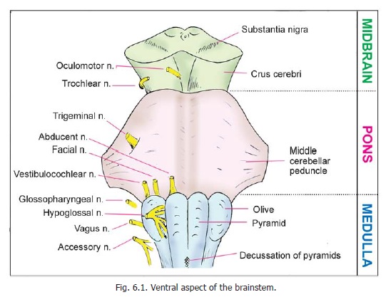
When the midbrain is viewed from the anterior aspect, we see two large bundles of fibres, one on each side of the middle line. These are the crura of the midbrain. The crura are separated by a deep fissure. Near the pons the fissure is narrow, but broadens as the crura diverge to enter the corresponding cerebral hemispheres. The parts of the crura just below the cerebrum form the posterior boundary of a space called the interpeduncular fossa (Fig. 8.7). The oculomotor nerve emerges from the medial aspect of the crus (singular of crura) of the same side.
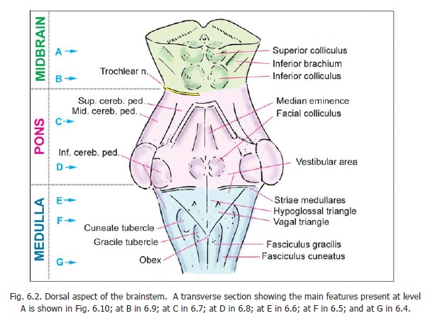
The posterior aspect of the midbrain is marked by four rounded swellings. These are the colliculi, one superior and one inferior on each side. Each colliculus is related laterally to a ridge called the brachium. The superior brachium (also called the superior quadrigeminal brachium, or brachiumof superior colliculus) connects the superior colliculus to the lateral geniculate body. Similarly, the inferior brachium (also called the inferior quadrigeminal brachium or brachium of inferior colliculus)connects the inferior colliculus to the medial geniculate body. Just below the colliculi, there is the uppermost part of a membrane, the superior medullary velum, which stretches between the two superior cerebellar peduncles, and helps to form the roof of the fourth ventricle. The trochlear nerve emerges from the velum, and then winds round the side of the midbrain to reach its ventral aspect.
In the description of the surface features of the brainstem, given above, reference has been made to the floor of the fourth ventricle.


Related Topics