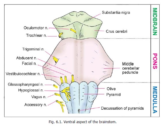
Chapter: Human Neuroanatomy(Fundamental and Clinical): Internal Structure of the Spinal Cord
Preliminary Review of the Internal Structure of the Brainstem
Preliminary Review of the Internal Structure of the Brainstem
The following description is confined to those features of internal structure that can be seen with the naked eye.
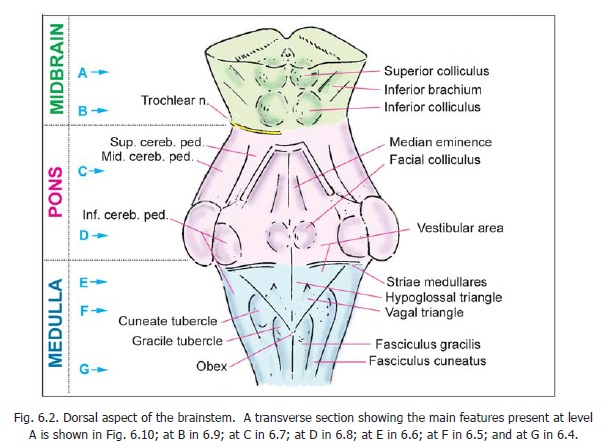
The main features of the internal structure of the brainstem are most easily reviewed by examining transverse sections at various levels. These are illustrated in Figs. 6.4 to 6.8. The levels represented in these figures are indicated in Fig. 6.2.
The Medulla
A section at the level of the pyramidal decussation (Fig. 6.4) shows some similarity to sections through the spinal cord. Thecentral canal is surrounded by central grey matter. The ventralgrey columns are present, but are separated from the central grey matter by decussating pyramidal fibres. The region behind the central grey matter is occupied by the fasciculus gracilis, medially;and by the fasciculus cuneatus laterally. Closely related to these fasciculi there are two tongue-shaped extensions of the central grey matter. The medial of these extensions is the nucleus gracilis, and the lateral is the nucleus cuneatus. More laterally, there is the spinal nucleus of the trigeminalnerve. When traced inferiorly, this nucleus reaches the second cervical segment of the spinal cord,where it becomes continuous with the substantia gelatinosa. Above, the nucleus extends as far as the
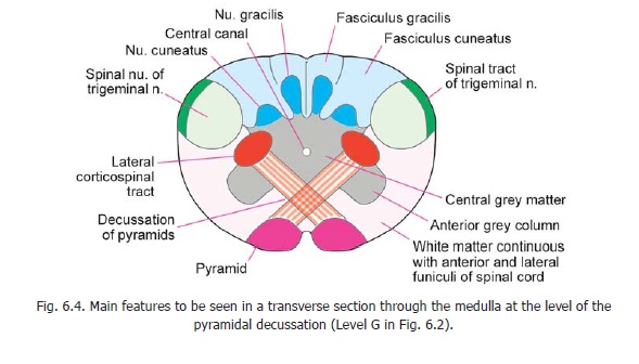
upper part of the pons. The spinal nucleus of the trigeminal nerve is related superficially to the spinaltract of the nerve. The ventral part of the medulla is occupied, on either side of the midline, by aprominent bundle of fibres: these fibres form the pyramid. The fibres of the pyramids are corticospinal fibres on their way from the cerebral cortex to the spinal cord. At this level in the medulla many of these fibres run backwards and medially to cross in the midline. These crossing fibres constitute the decussation of the pyramids. Having crossed the midline, the corticospinal fibres turn downwardsto enter the lateral white column of the spinal cord. The anterolateral region of the medulla is continuous with the anterior and lateral funiculi of the spinal cord.
A section through the medulla at a somewhat higher level is shown in Fig. 6.5. The central canal surrounded by central grey matter, the nucleus gracilis, the nucleus cuneatus, the spinal nucleus of the trigeminal nerve, and the pyramids occupy the same positions as at lower levels. The nucleus gracilis and the nucleus cuneatus are, however, much larger and are no longer continuous with the central grey matter. The fasciculus gracilis and the fasciculus cuneatus are less prominent. The region just behind the pyramids is occupied by a prominent bundle of fibres, the medial lemniscus, on
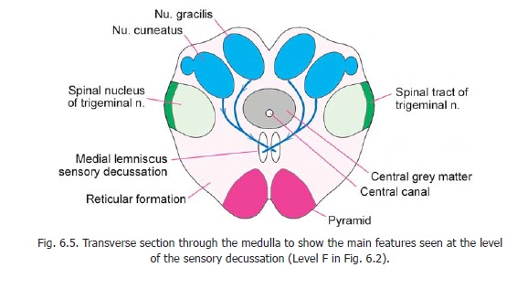
either side of the midline. The medial lemniscus is formed by fibres arising in the nucleus gracilis and the nucleus cuneatus. These fibres cross the midline and turn upwards in the lemniscus of the opposite side. The crossing fibres of the two sides constitute thesensory decussation. The region lateral to the medial lemniscus contains scattered neurons mixed with nerve fibres. This region is thereticularformation. More laterally there is a mass of white matter containing various tracts.
A section through the medulla at the level of the olive is shown in Fig. 6.6. The pyramids, the medial lemniscus, the spinal nucleus and tract of the trigeminal nerve, and the reticular formation are present in the same relative position as at lower levels. The medial lemniscus is, however, much more prominent and is somewhat expanded anteriorly. Lateral to the spinal nucleus (and tract) of the trigeminal nerve we see a large compact bundle of fibres. This is the inferior cerebellar peduncle which connects the medulla to the cerebellum. Posteriorly, the medulla forms the floor of the fourth ventricle. Here it is lined by a layer of grey matter in which are located several important cranial nerve nuclei.
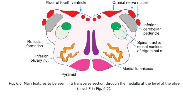
The inferior olivary nucleus forms a prominent feature in the anterolateral part of the medulla at this level. It is made up of a thin lamina of grey matter that is folded on itself like a crumpled purse. The nucleus has a hilum that is directed medially.
The Pons
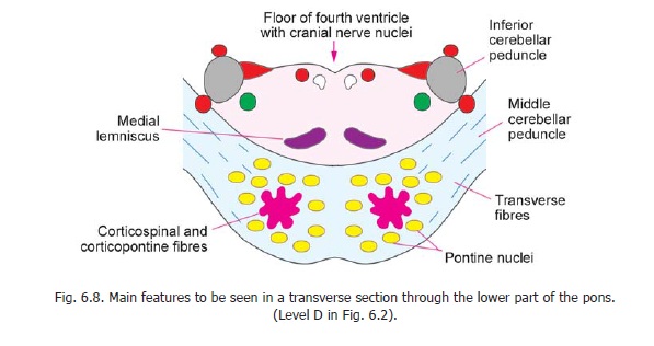
The pons is divisible into a ventral part and a dorsal part (Fig. 6.7).
The ventral (or basilar) part contains numerous transverse and vertical fibres. Amongst the fibres are groups of cells that constitute the pontine nuclei. When traced laterally the transverse fibres form the middle cerebellar peduncle. The vertical fibres are of two types. Some of them descend from the cerebral cortex to end in the pontine nuclei. Others are corticospinal fibres that descend through the pons into the medulla where they form the pyramids.
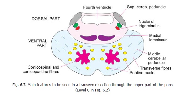
The dorsal part (or tegmentum) of the pons may be regarded as an upward continuation of the part of the medulla behind the pyramids. Superiorly, it is continuous with the tegmentum of the midbrain. It is bounded posteriorly by the fourth ventricle. Laterally, it is related to the superior cerebellarpeduncles in its upper part (Fig. 6.7); and to the inferior cerebellar peduncles in its lower part (Fig.6.8). The spinal nucleus and tract of the trigeminal nerve lie just medial to these peduncles. The medial lemniscus forms a transversely elongated band of fibres just behind the ventral part of the pons.
The Midbrain
For convenience of description, the midbrain may be divided as follows (Fig. 6.9).
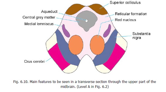
(a) The part lying behind a transverse line drawn through the cerebral aqueduct is called the tectum. It consists of the superior and inferior colliculi of the two sides.
(b) The part lying in front of the transverse line is made up of right and left halves called the cerebral peduncles. Each peduncle consists of three parts. From anterior to posterior side theseare the crus cerebri (or basis pedunculi), the substantia nigra and thetegmentum.
The crus cerebri consists of a large mass of vertically running fibres. These fibres descend from the cerebral cortex. Some of these pass through the midbrain to reach the pons, while others reach the spinal cord. The two crura are separated by a notch seen on the anterior aspect of the midbrain.
The substantia nigra is made up of pigmented grey matter and, therefore, appears dark in colour. The tegmentum of the two sides is continuous across the midline. It contains important masses of grey matter as well as fibre bundles. The largest of the nuclei is the red nucleus (Fig. 6.10) present in the upper half of the midbrain. The tegmentum also contains the reticular formation which is
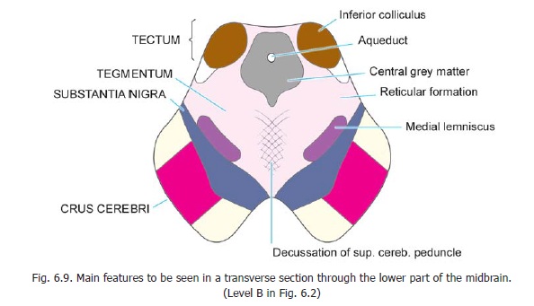
continuous below with that of the pons and medulla. The fibre bundles of the tegmentum include the medial lemniscus which lies just behind the substantia nigra, lateral to the red nucleus. The lowerpart of the tegmentum is traversed by a large number of fibres that cross the midline from one side to the other. These are the fibres of the superior cerebellar peduncles that have their origin in the cerebellum and decussate before ending in the red nucleus (and in some other centres).
It may be noted that some authorities describe the corresponding half of the tectum as part of the cerebral peduncle.
Related Topics