Chapter: Biochemistry: Living Cell
Sub cellular Organelles
Sub cellular Organelles
Cell Membrane
The cell is enveloped and thus separated from its
surroundings by a thin wall contains a rigid framework of polysaccharide chains
crosslinked with short peptide chains. Its outer surface is coated with
lipopolysaccharide. Cell membrane is also called as plasma membrane (or) plasma
lemma. The pili, not found in all bacteria have extensions of the cell wall.
The cell membrane contains about 45% lipid and 55% protein. The cell membrane
or plasma membrane have an average thickness of 75A°. The principal lipids are
phospholipids, sphingolipids and cholesterol. An important feature of these
lipids is they are composed of hydrophobic (water - insoluble) hydrocarbon
sections and hydrophilic (water soluble) units. The latter include charged
units (eg. phosphate or amino groups) and uncharged units (eg. hexose). In
water, such compounds orient themselves in such a way that only the hydrophilic
section is exposed to water. The hydrophobic components of individual molecules
tend to contact with other; this is accomplished either by arrangement into
micelles or by the formation of bilayers.
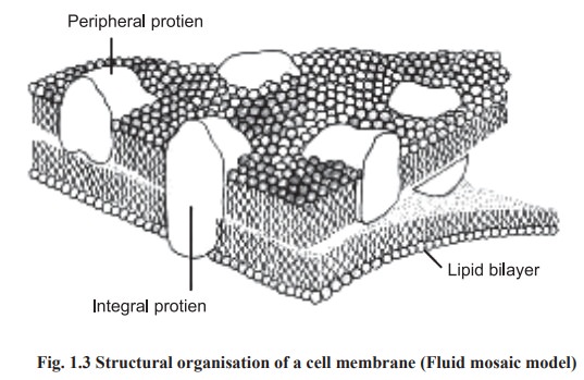
Two types of poteins are found in cell
membrane; viz. intrinsic or integral and extrinsic or peripheral (Fig.
1.3).Integral proteins are either partially or totally immersed in the lipid
bilayer and difficult to remove by any means other than the distruption of the
membrane with a detergent. Peripheral proteins are bound only to the surface of
membrane and interact only with the hydrophilic groups and therfore are readily
removed by extraction with an aqueous medium. The model of arrangement of
lipids and proteins in the memberane is known as fluid mosaic model.
Functions of cell membrane
·
The cell
membrane performs a number of important functions It holds the cell together
·
The membrane
is a selectively permeable boundary which allows water, certain required
nutrients and metal ions to pass freely It secrete waste products
·
It keeps out toxic materials
·
It
contain receptors to bind certain regulatory substances such as hormones which
regulate the various metabolic activities.
Cell Wall
Plant and bacterial cell membranes are
surrounded by a thick cell wall.
Bacterial cell wall
The bacterial cell is enclosed within a wall
that differs chemically from the cell wall of plants. The cell wall contains a
rigid framework of polysaccharide chain cross linked with short peptide chains
and its outer surface is coated with lipopolysaccharide. The pili, found in
some bacteria are extensions of the cell wall. In some bacteria the cell wall is
surrouned by an additional structure called a capsule.
The cell wall and capsule confer shape and form
of the bacteriam and also act as a physical barrier to the cell membrane. In
the absence of cell wall and capsule is mechanically fragile and the bacteria
would rupture.
Plant cell wall
The cell wall is a thick polysaccharide
containing structure immediately surrounding the plasma membrane. In
multicelllar plants, the plasma membrane of neighboring cells are separated by
these walls, and adjacent plant cell have their walls fused together by a layer
called the middle lamella. The cell
wall serves both as a protective and a supportive unit for the plant. The
degree to which the cell wall may be involved in the regulation of the exchange
of materials between the plant cell and its surroundings is difficult to assess
but is most likely restricted to macromolecules of considerable size. As in
animal cells,most of the regulation of exchanges between the cytoplasm and the
extracellular surrounding of plant cells is a function of the plasma membrane
Functions
The cell wall protects bateria against swelling
in hypotonic media. It is porous and allows most small molecules to pass. Some
of the pili are hollow and serve to transfer DNA from sexual conjugation.
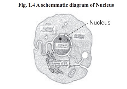
Nucleus
Nucleus is the heaviest particulate component
of the cell. Except matured mammalian erythrocytes, nucleus is found in almost
all cells. The nucleus about 4-6µm in diameter is surrounded by a perinuclear
envelope. At various position the outer membrane of the envelope fuses with the
inner membrane to form pores (Fig. 1.4). Nuclear pores provide continuity
between the cytosol and the contents of the nucleus (nucleoplasm). The electron
microscope reveals that the nuclear content consist of granular or fibrillar
structures. The nucleolus, a discrete body within the nucleus, contains
ribonucleic acids (RNA). The most important component of the nucleus is an
organised clumps of threadwork known as chromatin which is distributed
throughout the nucleus and contains most of the cellular deoxy ribonucleic
acids (DNA). Immediately before the cell division the chromatin organises into
simple thread like structures known as chromosomes which will eventually be
distributed equally to each daughter cell.
Functions
Take part in cell division Contain DNA molecules which are heriditary carriers.
Mitochondria
These are the largest particulate components of
the cytoplasm and represent upto 15% -20% of the dry weight of the cell. They
vary in shape (spherical, filamentous, sausage shaped) and size (0.5 to 3μ long
0.1 to 0.6μ wide).The number varies with the size and energy requirements of
the cell. For eg. flight muscles in birds contain rich amount of mitochondria
when compared to any other parts of the body
Electron microscopic studies show that a
mitochondrion has two membranes inner and outer which are separated from each
other by 50 to 100oA. The outer and inner membranes differ in lipid composition
and in enzyme content.
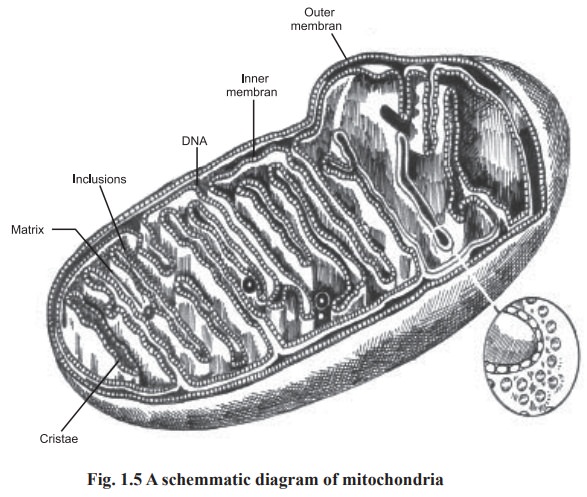
The inner membrane is very much folded to form
shelf - like structures of varying width. These shelf - like structures, known
as internal ridgs or cristae, extent into matrix of the mitochondrion
structure. Thus two structurally different space can be distinguished, the
intracristae space and the matrix space (Fig. 1.5). The matrix space is rich in
enzymes. The inner membrane shows the existance of knob like structures, which
are proteins involved in biological oxidations.
Functions
: The mitochondria are the ‘power houses’ of the
cell, where carbohydrates, lipids and
amino acids are oxidised to CO2 and H2O by molecular
oxygen, and the energy set free is stored in the form of adenosine triphosphate
(ATP). The enzymes involved in this energy conversion are located in the inner
membrane.
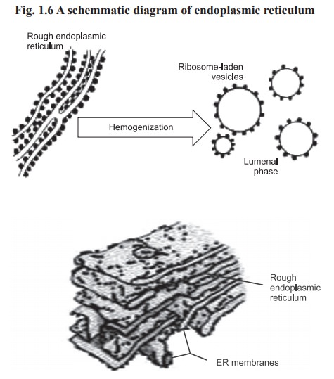
Endoplasmic reticulum
The endoplasmic reticulum consist of flattened
single membrane vesicles. These have the same lipid bilayer structure but
thinner than the cell membrane (about 7mm). The endoplasmic reticulum is of two
types; rough (RER) and smooth (SER). Only the rough type has small granules
known as ribosomes (Fig. 1.6).
Functions
RER is concerned with protein synthesis while
SER is concerned with lipids and glycoprotein synthesis. The cisternae
(enclosed spaces) of the endoplasmic reticulum play a role in the exchange of
material between the cell and the extra cellular fluid. The exchange of
material takes place by the process of pinocytosis.
Golgi apparatus
Golgi complex is a smooth membrane system
consists of flattened, single membrane vesicles which are often stacked (Fig.
1.7).
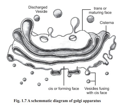
Functions
These are organelles to which the newly
synthesized proteins are transferred and temporarily stored. Small vesicles
arise peripherally by a pinching - off process. Some become vacuoles in which
secretory products are concentrated.
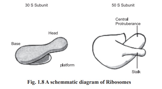
Ribosomes
The outer membrane of the endoplasmic reticulum
contain small granules commonly known as ribosomes, which are the smallest
particulate components of the cytoplasm. They are rich in ribonucleic acids.
Each ribosome has a large and a small subunit with a sedimentation constant of
50s and 30s respectively (Fig. 1.8). Each subunit contains about 65% RNA and
35% protein.
Functions
·
Ribosomes
are the sites of protein synthesis. Messenger RNA binds in the groove between
the subunits and specifies the sequence of amino acids in the growing
polypeptide chains. The proteins synthesized on membrane bound ribosomes must
pass successively through each of cytomembrane system.
·
Secretion
may involve the fusion of the vacuoles with the plasma membrane followed by a
discharge of the contents into the extra cellular space. This process is called
exocytosis.
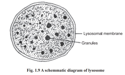
Lysosomes
Lysosomes are single membrane vesicles, having
intermediate size between microsomes and mitochondria. These are surrounded by
a lipoprotein membrane (Fig.1.9). Lysosomes are rich in many hydrolytic enzymes
such as phosphatase and ribonuclease and because of this, they are named as
lysosomes (Lyso means lytic action).
Functions
The hydrolytic enzymes of lysosome completely
destroy the foreign materials like pathogenic microorganism. They also serve to
digest cell components after cell death. Inside the macrophages these lysosomes
combine with vecuole which has engulfed the foreign particles and form
phagolysosomes. Inside these phagolysosomes foreign particles are degraded or
killed. The pathogen engulfed lysosomes are destroyed by the reticulo
endothelial system. Due to this action lysosomes are called as ‘Suicidal Bags’.
Peroxisomes
Peroxisomes are otherwise known as microbodies.
They are single - membrane vesicles of about 0.5 mm in diameter. They contain
catalase, D-amino acid oxidase, urate oxidase and other oxidative enzymes.
Functions
Microbodies participate in the oxidation of
certain nutrients. Hydrogen peroxide, the toxic reduction product of oxygen is
decomposed to form water in these organelles.
Cytoplasm
The intracellular cell content that posses both
soluble and insoluble constituents is called cytoplasm.
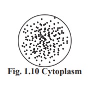
Cytosol
The soluble liquid portion of the cytoplasm is
known as cytosol in which the organelles are bathed. Cytosol is also known as
cell sap. Cell sap contains water, proteins, lipids and numerous other solutes
and is highly viscous (Fig.1.10).
Functions
Some important metabolic processes occur in the
cytosol are glycolysis, gluconeogenesis, activation of amino acids and
biosynthesis of fatty acids.
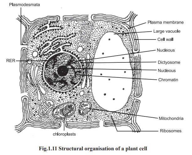
Plant Cells
Plant cells have cell wall made up of cellulose
and the cytoplasm consists of big vacuoles and chloroplasts (Fig.1.11).
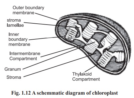
Chloroplasts
The ability to use light as a source of energy
for sugar synthesis from water and carbon dioxide is a special feature of
certain plant cells. This process, termed photosynthesis is carried out in
organelles called chloroplasts. These organelles are commonly ellipsoidal
structures bounded by an outer membrane but also containing a number of
internal membranes. Internally, the chloroplast consists of a series of
membranes arranged in parallel sheets called lamellae and supported in a
homogeneous matrix called the stroma. The membranes are arranged as thin sacs
called thylakoids that contain chlorophyll and may be stacked on top of one
another, forming structures called grana. Lamellar membranes connecting the
grana are called stroma lamellae (Fig.1.12).
Vacuoles
Although vacuoles are present in both animal
and plant cells, they are particularly large and abundant in plant cells, often
occupying a major portion of the cell volume and forcing the remaining
intracellular structures into a thin peripheral layer. These vacuoles are bound
by a single membrane and are formed by the coalescence of smaller vacuoles
during the plant’s growth and development. Vacuoles serve to expand the plant
cell without diluting its cytoplasm and also function as sites for the storage
of water and cell products or metabolic intermediates.
Related Topics