Chapter: Basic Radiology : Radiology of the Urinary Tract
Retrograde Pyelography/Cystography/Urethrography - Radiology of the Urinary Tract: Techniques and Normal Anatomy
Retrograde
Pyelography/Cystography/Urethrography
Direct injection of water-soluble
iodinated contrast material is a useful method of examining various regions of
the uri-nary tract. The advantage of this method of evaluation is the direct control
over the contrast injection rather than reliance on secondary excretion from
the kidney.
Retrograde pyelography, often
carried out in conjunction with cystoscopy, is performed by placing a small
catheter into the distal ureter. Contrast material is then injected through
this catheter into one or both ureters. Fluoroscopy and conven-tional
radiographs should then be obtained. This study usually results in excellent
evaluation of the ureter and intrarenal col-lecting system. The ureter is
typically seen in its entirety, which rarely occurs with other imaging studies.
Interpretation is sim-ilar to that of CT urography, with the caveat that the
contrast within the collecting system is under greater pressure than
physiologic conditions and mild ballooning of the calyces as well as occasional
extravasation can occur normally.
Imaging of the bladder is
performed with a cystogram, where a catheter is placed into the bladder and
contrast ma-terial is then injected. The contrast material is optimally
in-jected under fluoroscopic observation but occasionally is performed with
only static conventional radiographs, such as in the trauma setting. One
advantage to cystography is that vesicoureteral reflux can be evaluated during
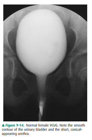
the conventional cystogram. The urethra may be evaluated with contrast material
via two methods. In one, the urethra is evaluated during voiding, often
following a cystogram (voiding cys-tourethrogram or VCUG). Alternatively, a
retrograde study may be performed (retrograde urethrogram). The urethra in the
male consists of four portions, including the prostatic, membranous, bulbous,
and penile portions. During voiding, the urethra is fairly uniformly distended
and tubular in ap-pearance. On a retrograde study, the more posterior urethra
(prostatic and membranous) is often contracted and seen as a thin wisp of
contrast. The female urethra appears as a short, slightly funnel-shaped tubular
structure during voiding (Figure 9-14). The urethra in males is generally
evaluated for injuries and strictures but may also be examined for filling
defects, masses, and fistulas.
Related Topics