Chapter: Basic Radiology : Radiology of the Urinary Tract
Nuclear Medicine - Radiology of the Urinary Tract: Techniques and Normal Anatomy
Nuclear Medicine
In general, the value of nuclear
imaging in the urinary tract is severalfold: functional information related to
the quantifiable collecteddata, lower radiation dose than traditional
radiographic tech-niques, and very low incidence of complications. Renal
eval-uation is typically performed by intravenous bolus injection of renal
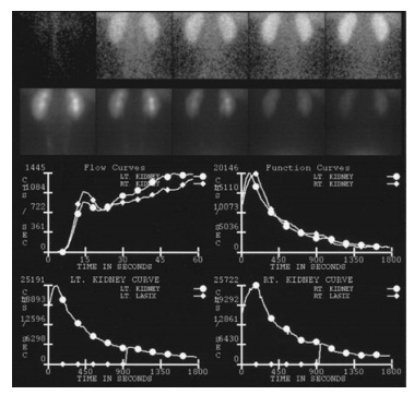
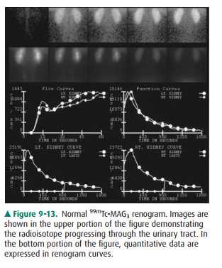
specific agents such as technetium-labeled mercap-toacetyltriglycine (Tc-MAG3).
Images are acquired every few seconds demonstrating renal blood flow with
additional im-ages obtained over several minutes showing renal uptake and
excretion. The recorded data can be used to produce images, but it also is
quantifiable and is employed to generate time-activity curves (Figure 9-13).
Information about renal perfu-sion, morphology, relative function of each
kidney, and excretion can be extremely useful in evaluation of conditions such
as renovascular hypertension, obstruction, and renal transplant examination.
Although anatomically oriented data can be obtained with other radioisotopes
that aggregate more in the renal parenchyma, in general, nuclear medicine renal
studies suffer from fairly low spatial resolution and are therefore often used
in conjunction with other imaging stud-ies. Radionuclide cystography is another
useful test used to diagnose and monitor vesicoureteral reflux. Here,
tech-netium pertechnetate is mixed with saline and infused into the bladder
with subsequent images obtained over the uri-nary tract. This study is quite
sensitive for the detection of significant reflux, but at a considerably lower
radiation dose than conventional cystography, making it particularly useful in
children, especially in those needing follow-up and repeated imaging. Another
important study is the radioactive iodine labeled metaiodobenzylguanidine
(MIBG) examina-tion. MIBG collects in adrenal medullary tissue and is useful in
diagnosis and evaluation of pheochromocytoma. Positron emission tomography
(PET) is evolving as a powerful imag-ing tool, especially when combined with CT
(PET/CT), com-bining the functional data of PET with the anatomic CT
information. Unfortunately, fluorine-labeled deoxyglucose (FDG), which is the
primary agent used in PET/CT, is nor-mally excreted by the kidneys, obscuring
urinary tract pathology and limiting utilization. PET/CT has, however, shown
promise in evaluation of possible metastatic disease.
Related Topics