Chapter: Human Nervous System and Sensory Organs : Brain Stem and Cranial Nerves
Reticular Formation

Reticular Formation
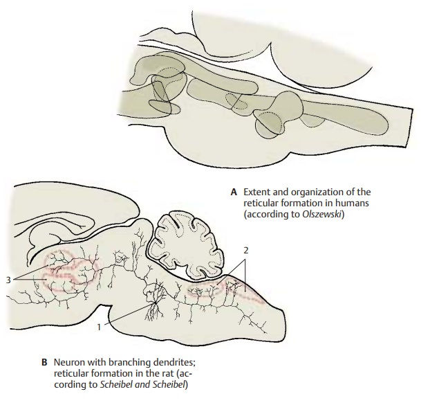
The scattered
neurons of the tegmentum and their network of processes form the reticular formation. This occupies the
centralarea of the tegmentum and expands from the medulla oblongata into the
rostral mid-brain. Several areas of different structure can be distinguished (A). In the medial part are magnocellular nuclei from where long ascending and descending fiber tracts originate.The
parvocellular lateral part is
regarded as an association area.
Many of
the neurons have long ascending or descending axons, or axons bifurcating into
an ascending and a descending branch. As shown by Golgi impregnation, such a
neu-ron (B1) can simultaneously
reach caudalcranial nerve nuclei (B2) and diencephalic nuclei (B3).
The reticular formation containsa large number of peptidergic neurons
(enkephalin, neurotensin, and others).
Afferent connections.The reticular forma-tion is
reached by impulses of all
sensorymodalities. Sensory spinoreticular fibersterminate in the medial
field of medulla ob-longata and pons, and so do secondary fibers of the
trigeminal and vestibular nu-clei. Collaterals of the lateral lemniscus bring
in acoustic impulses, while fibers of the tectoreticular fasciculus bring in
optic impulses. Experimental studies on stimula-tion have shown that reticular
neurons are excited more by sensory (pain), acoustic and vestibular stimuli
than by optic stimuli. Other afferent fibers originate from the cerebral
cortex, the cerebellum, the red nu-cleus, and the pallidum.
Efferent connections.The reticulospinal tract runs from the medialfield of medulla
oblongata and pons into the spinal cord. Bundles of the reticulothalamicfasciculus ascend to the intralaminar nucleiof the
thalamus (truncothalamus). Fiber bundles from the midbrain termi-nate in the oral
hypothalamus and in the septum.
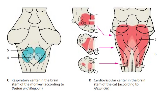
Respiratory and cardiovascular control centers. Groups
of neurons regulate respi-ration (C),
heart rate, and blood pressure (changes upon physical activity or emotion). The
neurons for inspiration are localized
in the central field of the lower portion of the medulla oblongata (C4), those for expiration are further dorsal and lateral (C5). The higher relay stations for inhibition and stimulation of
respiration lie in the pons (locus
ceruleus). The autonomic nuclei of the glossopharyngeal nerve and the vagus
nerve are involved in regulating heart rate and blood pressure (D). Electrical stimula-tion in the
caudal central field of the medulla oblongata causes a drop in blood pressure (depressor center) (D6), while elec-trical stimulation of the remaining reticular
formation in the medulla oblongata (D7)
leads to an increase in blood pressure.
Effect on the motor system.The
reticularformation has a differential effect on the spinal motor system. In the
medial field of the medulla oblongata lies an inhibition cen-ter; upon stimulation, the muscle tonedrops,
reflexes fail, and the electric stimula-tion of the motor cortex no longer
triggers a reaction. By contrast, the reticular forma-tion in pons and midbrain
has an enhancingeffect on the motor
system.
Ascending activation systems.The
reticu-lar formation has an effect on consciousness via connections to the
intralaminar nuclei of the thalamus. When strongly stimulated by sensory or
cortical input, the organism sud-denly becomes fully alert, a prerequisite for
attention and perception. Upon electrical stimulation of the reticular
formation, this wake-up function can
be objectively assessedby electroencephalography (EEG).
Related Topics