Chapter: Human Nervous System and Sensory Organs : Brain Stem and Cranial Nerves
Overview of Brain Stem and Cranial Nerves
Brain Stem and Cranial Nerves
Overview
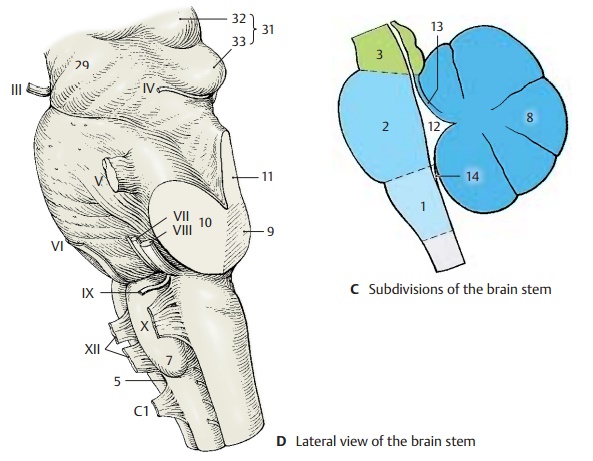
The brain stem, or encephalic trunk, is sub-divided into three sections: the medulla ob-longata (elongated spinal
cord) (C1), the pons (bridge) (C2), and
the mesencephalon (midbrain) (C3).
It is
this part of the brain that is underlain by the chorda dorsalis (notochord)
during embryonic development and from where ten pairs of genuine peripheral
nerves (cranial nerves III – XII) emerge. The cerebel-lum, which in ontogenetic terms alsobelongs to it, will be
discussed separately because of its special structure.
The medulla oblongata between the
pyra-midal decussation and the lower border of the pons represents the
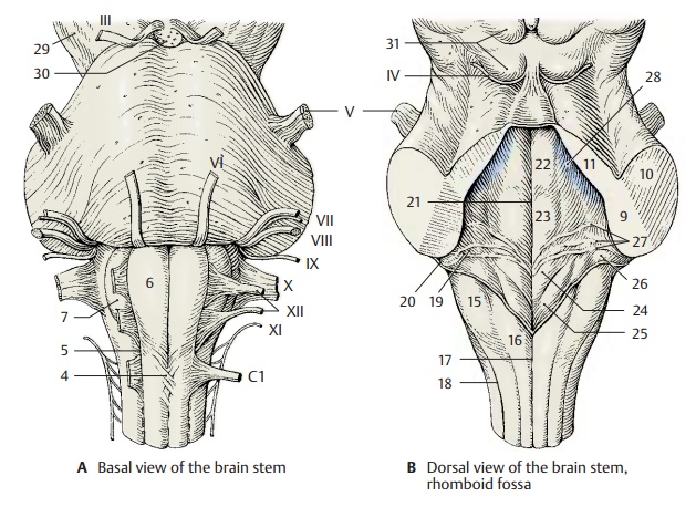
transition from the spinal cord to the brain. The anterior medianfissure, which is interrupted by the pyra-midal decussation (A4), and the antero-lateral sulcus (AD5)
on each side extend upto the pons. The anterior funiculi thicken below the pons
to form the pyramids (A6). Lateral to them on each side bulge
the olives (AD7).
The pons forms a broad arching bulge with
prominent transverse fibers. Here, de-scending pathways from the brain are
re-layed to neurons extending to the cerebel-lum.
The
posterior surface of the brain stem is covered by the cerebellum (C8). Upon
its removal, the cerebellar peduncles
are cut through on both sides, namely, the inferiorcerebellar
peduncle (or restiform body)(BD9), the middle cerebellar peduncle (or brachium
pontis) (BD10), and the superior cerebellar peduncle (or brachium conjunc-tivum) (BD11). Removal of the cerebellumopens
the fourth ventricle (C12), the tent-shaped roof of which is
formed by the supe-rior medullary velum (C13) and the inferior medullary velum (C14).
The floor of thefourth ventricle, the rhomboid
fossa, thus becomes exposed (B).
Medulla oblongata and pons together form the hindbrain, also known as rhombencephalon, named after this
fossa. The posterior funiculi (see p. 56)thicken on both sides to form the tubercle ofthe cuneate nucleus (B15) and the tubercle of the gracile nucleus (B16); they are borderedby the posterior
median sulcus (B17) and by the posterolateral sulcus on each side (B18).
The fourth ventricle forms on each side the
lateral recess (B19) which opens to the sub-arachnoid space by the lateral aperture(foramen of Luschka) (B20).
An unpaired opening lies below the inferior medullary velum, the median aperture (foramen ofMagendie). The floor of therhomboid fossa shows bulges
near the me-dian sulcus (B21); they are caused by cranialnerve
nuclei, namely, the medial eminence(B22), the facial colliculus (B23),
the trigonof the hypoglossal nerve (B24), the trigon of the vagus nerve (B25),
and the vestibular area (B26). The rhomboid fossa is crossed
bymyelinated nerve fibers, the medullarystriae
(B27). The pigmented nerve cells
ofthe locus ceruleus (B28) shine blueish through the floor of
the rhomboid fossa. They are mostly noradrenergic and project into the
hypothalamus, the limbic system, and the neocortex. The locus ceruleus also
contains peptidergic neurons (enkephalin, neurotensin).
The
anterior surface of the midbrain, or
mesencephalon, is formed by thecerebralpeduncles (A29) (descending cerebral path-ways). Between them lies the interpeduncu-lar fossa (A30); its floor is perforated
bynumerous vessels and is known as the pos-terior
perforated substance. At the posteriorsurface of the midbrain lies the tectal plate(or quadrigeminal plate) (BD31)
with two upper elevations, superior
colliculi (D32), the relay
station of the optic system, and two lower elevations, the inferior colliculi (D33),
the relay station of the acoustic sys-tem.

Related Topics