Chapter: Human Nervous System and Sensory Organs : Brain Stem and Cranial Nerves
Facial Nerve - Cranial Nerves (V, VII - XII)

Facial Nerve
The seventh cranial nerve supplies motor
fibers to the muscles of facial expression; in a nerve bundle emerging
separately from the brain stem, called the intermediate nerve, it carries taste
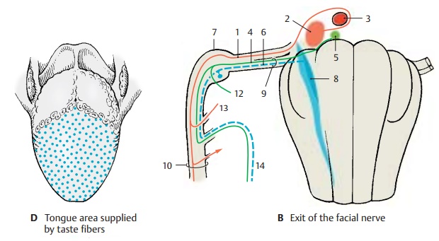
fibers and viscero-efferent secretory (parasympathetic) fibers. The motor fibers (AB1) originate from the large, multipolar neurons in the nucleus ofthe facial nerve (AB2). They arch aroundthe abducens nucleus (AB3) (internal genu ofthe
facial nerve) and emerge on the lateralaspect of the medulla oblongata from
the lower border of the pons. The cells of the preganglionic secretory fibers (AB4) form the superior
salivatory nucleus (AB5). The taste fibers (AB6) originate from the pseudo-unipolar cells in the geniculate ganglion (BC7) and terminate in the cranial
section of the solitary nucleus (AB8). The visceroefferentand taste
fibers do not arch around the ab-ducens nucleus but join the ascending limb of
the nerve and emerge as intermediatenerve
(B9) between the facial nerve
and thevestibulocochlear nerve.

Both
parts of the nerve pass through the inner auditory canal, the internal acousticmeatus (petrous part of
temporal bone, in-ternal acoustic pore, see vol. 1), and enter the facial canal as a nerve trunk. At the
bend of the nerve in the petrous bone (externalgenu
of the facial nerve) lies the
geniculate ganglion (BC7). The
canal continues abovethe tympanic cavity and turns caudally toward the stylomastoid foramen (BC10), through which the nerve leaves
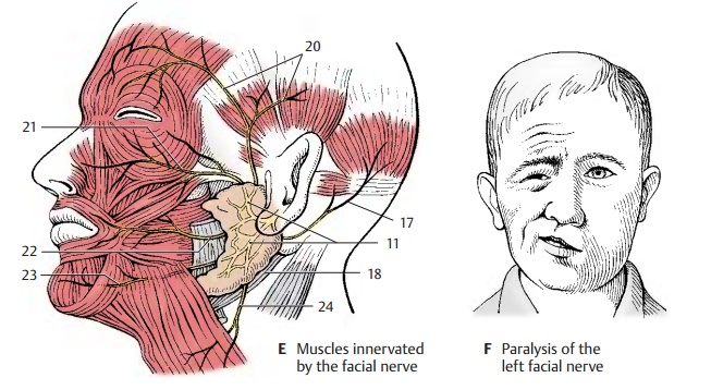
the skull. The nerve ramifies into terminal branches (parotid plexus) (E11) in
the parotidgland.
The
greater petrosal nerve (BC12), the
stapedius nerve (BC13), and the
chorda tympani (BC14) branch off
inside the facialcanal. Thegreater petrosal nerve(pregan-glionic
secretory fibers for the lacrimal gland, nasal glands, and palatal glands)
originates from the geniculate ganglion, ex-tends through the hiatus for the lesserpetrosal nerve into
the cranial cavity andover the anterior aspect of the petrous bone through the foramen lacerum and finallythrough the pterygoid canal to the pterygo-palatine ganglion (C15). Thestapedius nervesupplies the stapedius muscle in the middle ear. The
chorda tympani (BC14) branches off above the stylomastoid foramen, runs beneath the
mucosa through the tympanic cavity and further to the petrotympanic fissure,
and finally joins the lingual nerve (C16). It contains taste fibersfor the
anterior two-thirds of the tongue (D)
and preganglionic fibers for the subman-dibular and sublingual glands as well
as various lingual glands.
Before
it enters the parotid gland, the facial nerve gives off the posterior auricular nerve (E17) as well as branches to the
posterior belly of the digastric muscle
(CE18) and to the stylohyoid muscle (C19). The parotid plexus gives off the temporal branches (E20),
the zygomatic branches (E21), the buccalbranches (E22),
the marginal mandibular branch (E23), and the cervical branch (E24)for
the platysma . The
branches provide innervation to all the muscles of fa-cial expression.
Ramifications
of the cervical branch lying beneath the platysma form the superficialcervical ansa by anastomosing withbranches of
the sensory transverse cervical nerve. The small branches departing from the
ansa are mixed sen-sorimotor nerves. The terminal ramifica-tions of temporal
branches, buccal branches, and marginal mandibular branch form similar plexuses
with branches of the trigeminal nerve.
Clinical Note: Injury to the nerve results inatony of all muscles of the
affected half of the face. The mouth region drops, and the eye can no longer
close (F). There is increased
sensitivity to sound, hyperacusis.
C25 Trigeminal ganglion.
Related Topics