Chapter: Human Nervous System and Sensory Organs : Brain Stem and Cranial Nerves
Base of the Skull
Base of the Skull
The base of the skull holds the
brain. Three bony depressions on each side correspond to the basal aspects of the
brain; the basal aspect of the frontal lobe lies in the anteriorcranial fossa (A1),
that of the temporal lobe inthe middle
cranial fossa (A2), and the
basal aspect of the cerebellum in the posteriorcranial
fossa (A3). (For the
participation ofbones in and the boundaries of the cranial fossae, see vol. 1.)
The cranial cavity is lined by a hard meninx, the dura mater; its two layers form both a cover for the brain and the
periosteum. Embedded between these two layers are large venous sinuses. Nerves
and blood vessels pass through numerous foramina in the base of the skull (see
vol. 1).
On the floor of the anterior
cranial fossa close to the midline, the olfactory
nerves pass through the openings of the thin lamina cribrosa to the
olfactory bulb (A4).The sella turcica rises between the two
middle cranial fossae; its depression con-tains the hypophysis (A5), which
is attached to the floor of the diencephalon. Lateral to the sella turcica, the
internal carotid artery
(A6) passes through the carotid
canal into the cranial cavity. Its S-shaped course by-passes the cavernous sinus (A7). The opticnerve (A8) enters the cranial cavity
throughthe optic canal in the medial area of the fossa, while the eye-muscle
nerves leave the cavity through the superior orbital fissure (see vol. 1). The
paths of the abducens nerve (A9) and the trochlear nerve (A10)
are charac-terized by their intradural position. The ab-ducens nerve enters the
dura at the middle level of the clivus, and the trochlear nerve enters at the
edge of the clivus at the attach-ment of the tentorium. The oculomotor nerve (A11) and the trochlear nerve run through the lateral wall of the
cavernous sinus, and the abducens nerve through the laterobasal sinus of the
internal carotid artery (see vol. 2). The trigeminal
nerve (A12) reaches below a
dural bridge into the middle cranial fossa where the trigeminal ganglion (A13)
lies in a pocket formed by the two dural layers, the trigeminal cavity. The three trigeminalbranches leave the cranial
cavity through different openings; after passing through the wall of the
cavernous sinus, the ophthal-mic nerve (A14) extends with its branchesthrough
the orbital fissure, the maxillarynerve (A15) through theround foramen,
andthe mandibular nerve (A16) through the ovalforamen.
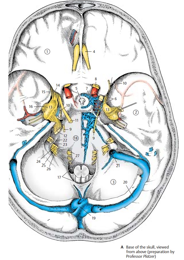
The two posterior cranial fossae
surround the foramen magnum (A17) to which the clivus (A18) descends
steeply from the sellaturcica. The brain stem rests on the clivus, and the
cerebellar hemispheres fit into the two basal fossae. From the confluence ofsinuses (A19), the transverse sinus (A20)
em-braces the posterior cranial fossa and opens into the internal jugular vein (A21).
The facialnerve (A22) and the vestibulocochlear nerve (A23)
enter the auditory canal, the internalacoustic
meatus. Basal to the meatus, theglossopharyngeal
nerve (A24), vagus nerve (A25), and accessory nerve (A26) pass through the anterior part of the jugularfossa. The fiber bundles of thehypoglossalnerve (A27)
pass as a single nerve throughthe hypoglossal
canal.
Related Topics