Chapter: Medical Electronics : Bio-Chemical and Non Electrical Parameter Measurement
Respiratory Rate Measurement
RESPIRATORY RATE MEASUREMENT
Respiratory
system provides a means of acquiring oxygen and eliminating CO2.
Various laws are involved in the understanding of respiratory functions.
Various
Gas laws are given below:
1. BOYLE’S LAW: It states that at constant
temperature, the volume of gas varies inversely
with the pressure.
V2/V1
=P1/P2 here temperature T= constant
V2=
Final volume
V1 = Initial
volume
P1 = Original (initial) pressure
P2 = Final
pressure
2. CHARLE’S LAW: It states that, at
constant pressure, the volume of gas isdirectly proportional to the absolute
temperature.
V2/V1
=T2/T1 Here pressure P=constant
V2,
V1 =Final, initial volume
T1
=original temperature
T2
= Final temperature
3 . HENRY’S LAW : It states that, if the
temperature is constant, the quantity of a gas that goes into a solution is
directly proportional to the partial pressure of that gas . The gas with the
greater partial pressure will have more mass in solution.
4. DALTON’S LAW :
It states
that, the total pressure exerted by a mixture of gases is equal to the sum of
the partial pressures of various gases.
PT
=P1 + P2 + …………… +Pn
PT
= total pressure
P1,
P2 ,P3 = partial pressure of various gases
TYPES OF RESPIRATION
Respiration is nothing but the interchange of
gases between an organism and the living medium Internal respiration is the exchange of gases between the blood
stream and nearby cells
.External respiration is the exchange of gases between the lungs
and blood stream .
Lungs Volumes and Capacities (Respiration
Parameters) Or (LVC)
Respiration
parameters are used to indicate the state of respiratory function , including
lung volumes and capacities , airway resistance , lung compliance , etc .
Dead Air
Only a
portion of the air entering the respiratory system actually reaches the alveoli
. The volume of air that is not available for gas exchange with the blood is
known as dead air . The total dead space is less less than 30 percentage of the
total volume .
Tidal Volume (TV)
Tidal volume
is the depth of breathing or the volume of gas inspired or expired during each
respiratory cycle. It is equal to 500 ml for a normal person .
Inspiratory Reserve Volume (IRV)
It is the
maximal amount of gas that can be inspired from the end- inspiratory position (
Extra inspiration from the high peak tidal volume . It is equal to 3600 ml for
a normal person
Expiratory reserve volume (ERV)
It is the
maximal amount of gas that can be end expiratory level. It is equal to 1200 ml.
Residual Volume(RV)
It is the
amount of gas remaining in the lungs at the end of maximal expiration. It is
equal to 1200 ml.
Minute Volume (MV)
It is the
volume of air breathed normally for 1 minute.
Total Lung Capacity(TLC)
It is the
amount of gas contained in the lungs at the end of maximal inspiration and it
is the sum of inspiratory capacity(IC) and functional residual capacity (FRC).
TLC is of 6000 ml for a normal person.
Vital Capacity(VC)
It is the
maximum amount of gas that can be expelled from the lungs by forceful effort
from maximal inspiration. It is 4800 ml for a normal person.
Inspiratory Capacity(IC)
It is the
maximum amount of gas that can be inspired from the resting expiratory level
and it is the sum of tidal volume and inspiratory reserve volume. It is equal
to 3600 ml for a normal person.
Functional Residual Capacity(FRC)
It is the amount of gas remaining in the lungs at
the resting expiratory level. FRC = ERV + RV
Airway resistance
It
relates to the ease with air flows through tubular respiratory structures. In
smaller tubes, airway resistance is high.
Lung Compliance
It is the
ability of the alveoli and lung tissue to expand on inspiration.
Lung Elasticity
It is the
ability of the lung’s elastic tissues to recoil during expiration
Intra thoractic Pressure
It is the
positive and negative pressure occur within the thoracic cavity Types of
respiration rate measurement
1. Displacement
method
2. Thermistor
method
3. Impedance
pneumography
4. CO2
method
5. Apnoea
detectors
Displacement Method
In this
method the transducer is hold by an elastic band which goes around the
chest.The respiratory movements results in a corresponding resistance changes
of the strain gauge. It is connected as one arm of a wheatstone bridge circuit.
Its output varies with chest expansion. This output corresponds to the
respiration activity.
Thermistor Method
Generally
there is a temperature difference between inspired and expired air. This
temperature is sensed by placing thermistor in front of nostrils. Thermistor is
placed by using suitable stand. The thermistor is connected with the bridge
circuit. So unbalance signal is amplified to get the respiratory activity.
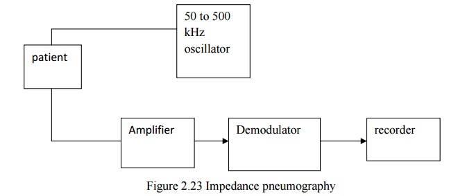
1. IMPEDANCE PNEUMOGRAPHY
This is
the indirect method of measurement . impedance pneumography is based on the
fact that , the a.c impedance across the chest of a patient changes as
respiration occurs . 50-50KHz a.c signal is produced by oscillator circuit and
is given to the chest of the patient through electrodes.
The
signal voltage applied to the amplifier (Differential amplifier) block is the
voltage drop across the resistance .
V =
I(R+ R)
Where V=
Output voltage (V)
I=
Current through the chest (A)
R= chest
impedance without respiration (R)
R= change
of chest impedance due to respiration (Q)
The output
of the amplifier is given to demodulator and filter block. Hence low pass
filter is used to remove the residual carrier signal. The output of the
impedance pneumograph contains respirating rate data.
CO2 Method
Respiration
rate can be measured by measuring CO2 in expired air. This CO2
method of measurement is based on the absorption property of infrared rays by
certain gases .When infrared rays are passed through the expired air which
contains certain amount of CO2, some of radiations are absorbed by
it. So, there is loss of heat energy associated with the rays .The detector
changes the loss in heating effect of the rays into an electrical signal. It is
used to get the average respiration rate. Two infrared sources are available in
this set up. The beam from one infrared source falls on the test cuvette side.
The beam from another infrared source falls on the reference cuvette side.
The
detector has two identical portions. These portions are separated by a thin,
flexible metal diaphragm. The detector is filled with a sample of pure CO2.
Because of the absorption of CO2 in the test cuvette. The beam
falling on the test side of the detector is weaker that falling on the
reference side. The gas in reference side is heated more than that on the test
side. So diaphragm is pushed slightly to the test side of the detector. This
diaphragm forms one plate f a capacitor. The a.c signal appears across the
detector is amplified and recorded using recorder. The amplified output is
integrated. It is used for continuous monitoring the respiration rate.
Apnoea Detectors
Apnoea is
the stopping of breathing. It leads to the arrest of the circulation. It can be
occurred at the conditions like head injury, drug overdose, etc. It can also
occur in premature babies during the first week of life because of their
immature nervous system. If apnoea persists for a prolonged period, then brain
functions can be severly damaged. So apnoea patients are closely monitored.
Apnoea monitor is used to watch the apnoea patients respiration rate. Apnoea
monitor gives audio signals and visual signals, when no inspiration occurs for
a particular period of time. Input from the sensor is connected with the
amplifier circuit having high input impedance.
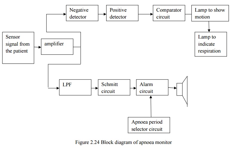
The output of the amplifier circuit is connected
with motion and respiration channel blocks. Motion channel block differentiates
motion and the respiration based on the frequency. The frequency below 1.5 Hz
is identified as respiration. The frequency above 1.5 Hz is identified as
motion. High frequency signal above the threshold is sensed by positive
detector.
The
frequency below the threshold is sensed by negative detector. The output of the
motion channel is connected with comparator circuit. It compares the amplitude
of motion and respiration. Based on the output corresponding lamp will glow. In
the respiration channel, low pass filter is used to remove high frequency
signal. If there is no respiration, then schmid trigger circuit gives the
output to switch on the alarm.
Apnoea
period selector circuits contain low frequency alarm oscillator and tone
oscillator, and audio amplifier. Apnoea perriod selector circuit drives the
alarm circuit. The output of alarm circuit is connected with the sp eaker. So,
where there is no respiration for a period of 10 or 20 sec, then audio signal
through the speaker and visual signal through the flash light is delivered.
Related Topics