Chapter: Human Nervous System and Sensory Organs : The Ear
Outer Ear - Structure of The Ear

Outer Ear
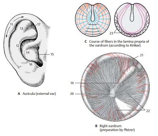
The auricle, or pinna (A, D1), with the ex-ception of the lobule,
contains a scaffold of elastic cartilage. The shapes of the auricular
projections and depressions are different in each person and are genetically
determined. The shapes of the following parts are in-herited: helix (A11), antihelix (A12), scapha (A13),
concha (A14), tragus (A15), antitragus (A16), and triangular fossa (A17).
In the past, the features of the auricle were of special importance for
establishing paternity.
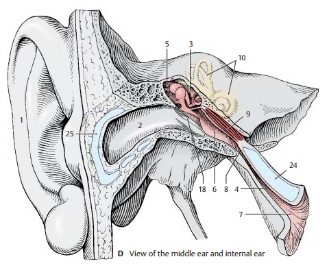
The
entrance of the outer ear canal, or ex-ternal acoustic meatus (D2), is formed by agroovelike
continuation of the auricular car-tilage (D25)
and is completed with connec-tive tissue to form a uniform passage. The passage
is lined with epidermis, and large ceruminous
glands lie beneath the epidermis.

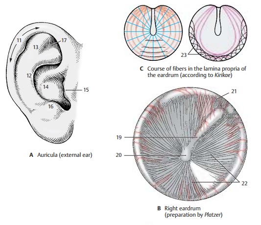
The
outer ear canal ends with the eardrum,
or tympanic membrane (B, D18),
which is obliquely placed in the meatus. When viewed from the outside, the mallearstria (B19) can be recognized; it is caused by the attachment of the
manubrium of the mal-leus, which reaches to the umbo of the tym-panic membrane (B20), the innermost pointof the funnel-shaped eardrum. Above the
upper end of the mallearstria (mallearprominence)
lies a lax, thin part of the ear-drum, the reddish pars flaccida (B21),
which is separated from the firm, gray and shiny pars tensa(B22) by two
mallear folds. Theeardrum is covered externally by skin and internally by a
mucosa. Between these lies the lamina propria of the pars tensa; it con-tains
radial and nonradial fibers (C). The
lat-terare circular, parabolic, and transversal. The fibrocartilaginous annulus (C23)
forms the anchoring tissue of the eardrum.

Related Topics