Chapter: Human Nervous System and Sensory Organs : The Ear
Middle Ear - Structure of The Ear

Middle Ear
Tympanic Cavity (A, B, D)
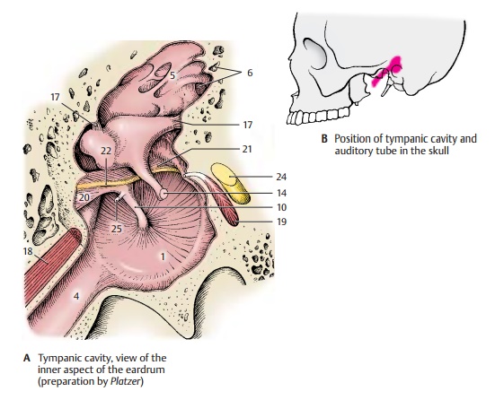
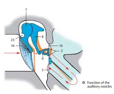
The tympanic cavity is a narrow, vertical space with the eardrum (AD1) obliquely placed in its lateral wall. Its medial wall has two openings leading to the internal ear, namely, the oval window, or vestibular win-dow(D2), and theround window, or cochlear window (D3). The roof of the tympanic cav-ity, the tegmental wall, is relatively thin and borders on the surface of the petrous pyra-mid. The floor of the tympanic cavity is formed by a thin layer of bone; beneath it runs the jugular vein.
The tympanic cavity continues in anterior direction as the auditory tube (A4). Its superior part opens posteriorly into the mastoid antrum (A5), a round space into which numerous small cavities open, the mastoid air cells (A6). These air-containingcavities are lined with mucosa and form a system of chambers that penetrates the en-tire mastoid bone; they may even extend into the petrous bone.
Auditory Ossicles (A, C, D)
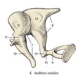
The three auditory ossicles, or ear bones, form together with the eardrum the sound-conducting apparatus. They are called the hammer, ormalleus(CD7), the anvil, orincus(CD8), and the stirrup, or stapes (CD9). The manubrium of the malleus (ACD10) is firmlyattached to the eardrum and connected via its neck (C11) to the head of the malleus (C12). The malleus has a saddle-shaped ar-ticular surface that contacts the body of theincus (C13). Its lenticular process (AC14),which is attached to the long limb of the incus and projects at a right angle, carries the articular surface for the head of thestapes (C15). The footplate of the stapescloses the vestibular window; it is attached at the margin by the annular ligament of the stapes (D16). Several ligaments (A17) con-nected to the wall of the tympanic cavity keep the ossicles in place.
The auditory ossicles transmit to the inner ear the vibration of the eardrum caused by sound waves. For this purpose, malleus andincus act like an angular lever, and the stapes performs a tilting movement. The footplate of the stapes transmits the vibra-tion to the fluid of the inner ear. The move-ment of the fluid through the cochlea is simplified in diagram (D); in reality, it takes a spiral course inside the cochlea. Tension in the system is controlled by two muscles with antagonistic effects, namely, the tensor tympani muscle (A18) and the stapedius muscle (A19).
The mucosa lining the tympanic cavity and covering the auditory ossicles forms various folds, such as the anterior (A20) and poste-riormallear folds (A21) which envelope the chorda tympani (A22). The folds form several mucosal pouches. The superiorrecess of the tympanic membrane, Prussak’spouch (D23), lies between the pars flaccida of the eardrum and the neck of the malleus; it plays an important role in ear infections.
A24 Facial nerve.
A25 Tendon of the tensor tympani muscle.
Medial Wall of the Tympanic Cavity (A – C)
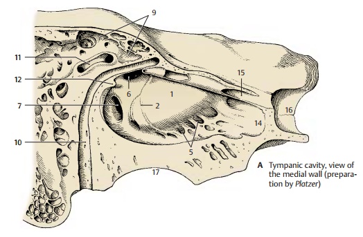
The medial wall, or labyrinthic wall, sepa-rates the tympanic cavity from the internal ear. The prominence in its middle region, the promontory of the tympanic cavity (A1), is caused by the basal convolution of the cochlea. In a bifurcated groove, the sulcus ofthe promontory (A2), lies the tympanic plexus (C3); it is formed by the tympanic nerve (C4) of the glossopharyngeal nerveand by the sympathetic fibers of the carotid plexus of the internal carotid artery. The promontory is anteriorly delimited by the tympanic cells (A5). In the medial wall, the oval window,vestibular window(A6), and the round window,cochlear window(A7), openinto the inner ear. The stapes (C8) rests in the vestibular window and closes it with its foot plate. The cochlear window is closed by the secondary tympanic membrane. In the posterior wall opening to the mastoid an-trum(A9) run two arched canals, the facial canal (A10) and the lateral semicircular canal (A11); both canals cause protrusionson the wall of the tympanic cavity, namely, the prominence of the facial canal and the prominence of the lateral semicircular canal.A bony protrusion, the pyramidal eminence (A12), contains an opening at its tip through which the tendon of the stapedius muscle (C13) enters. In anterior direction, the tym-panic cavity leads into the semicanal of theauditory tube (A14). Above it lies the semi-canal of the tensor tympani muscle (A15).The two semicanals are incompletely sepa-rated by a bony septum and together form the musculotubal canal. The medial wall at the level of the opening of the tympanic tube (carotid wall) separates the tympanic cavity from the carotid canal (A16); the bony floor (jugular wall) separates it from the jugular fossa (A17). Also shown are thejugu-lar vein (B18) and the internal carotid artery(B19).
Clinical Note: The osseous roof and floor ofthe tympanic cavity may be very thin. In case of purulent otitis media, the infection can penetrate either through the roof and progress to meninges and brain (meningitis, cerebral abscess in thetemporal lobe), or through the floor and progressto the internal jugular vein (jugular thrombosis)
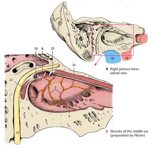
Muscles of the Tympanic Cavity (C)
The tensor tympani muscle (C20) origi-nates from the cartilaginous wall of the tympanic tube and from the osseous wall of the canal. Its narrow tendon bends away from the cochleariform process (C21) and in-serts on the manubrium of malleus. The muscle is innervated by the tensor tympaninerve of the mandibular nerve. Thestape-dius muscle (C22) originates in a smallbony canal which usually communicates with the facial canal. Its small tendon passes through the opening of the pyramidal emi-nence and inserts at the head of the stapes. The muscle is innervated by the stapediusnerve of the facial nerve (C23).
The two muscles control the tension of thesound-conducting apparatus. The tensor tym-pani muscle pulls the eardrum inward and pushes the footplate of the stapes into the vestibular window, thus increasing the sen-sitivity of the transmission. The stapedius muscle levers the footplate of the stapes out of the vestibular window and thus dampens the transmission. Hence, the two muscles are antagonists.
Clinical Note: Paralysis of the facial nervecauses loss of function in the stapedius muscle and deficient dampening of sounds; the patients suffer from hyperacusis, an increased sensitivity to sound.
Related Topics