Chapter: Human Nervous System and Sensory Organs : The Ear
Organ of Corti - Structure of The Ear
Organ of Corti
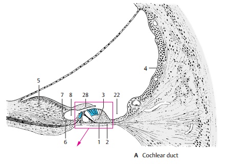
The basilar membrane (AB1) is covered on its lower surface by connective tissue cells
that line the scala tympani (AB2);
on its upper surface rests the organ of
Corti, or spiralorgan (A3,
B). Lateral to the spiral organ, theepithelium continues as the vascular stria (A4), which contains numerous intra-epithelial capillaries. Medial
to the spiral organ, at the margin of the bony spiral lamina, lies a thickened
tissue layer derived from the inner periosteum, the limbus of thespiral lamina (A5);
it is covered by epithelialcells. The limbus tapers off into two lips, the tympanic lip of the limbus(A6) and thevesti-bular lip of the limbus(A7),
which enclosethe internal spiral sulcus
(A8).
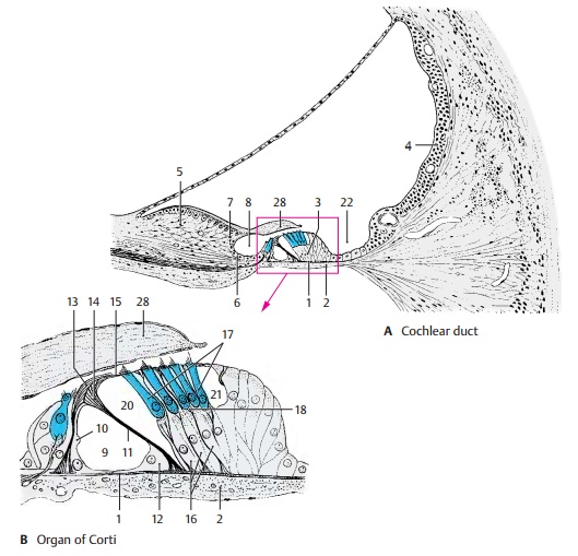
The
organ of Corti extends in a spiral from the basal convolution to the cupula of
the cochlea. The figures (A, B) show cross sec-tions through the
organ; it consists of sensory cells and a variety of supporting cells. The
center is occupied by the innertunnel (B9), which contains a perilymph-like
fluid, the cortilymph. Its medial wall is formed by the inner pillar cells (B10)
and its lateral wall by the obliquely arranged outerpillar cells (B11).
The pillar cells have a widebase (B12)
containing the cell nucleus, a narrow middle part, and a head. They con-tain
long bundles of supporting tonofila-ments. The inner pillar cell forms the head
plate (B13), while the outer pillar
cell forms a round head part (B14),
which approaches the head plate from below, as well as a flat process, the phalangeal process (B15). Later-ally follows the group of Deiters’ supportingcells (outer phalangeal cells) (B16); thesecarry the sensory cells (B17, C) on a protru-sion of their lower part. Their tonofilament bundles
branch below the sensory cells to form a support
basket (B18). From each Deiters’
cell, a narrow process ascends be-tween the sensory cells and ends with a flat
phalangeal process (C19). The
phalangeal processes together form a perforated super-ficial membrane, the reticular membrane, and the apical ends
of the sensory cells are firmly secured in the openings of the mem-brane.
Between the outer pillar cells and the Deiters’ cells lies the inner tunnel, or Corti’sspace (B20), and
laterally to the Deiters’cells lies the small outer tunnel, or Nuel’sspace (B21). This is followed by a group
ofsimple, elongated supporting cells that merge into the epithelium of the
vascular stria to form the medial wall of the externalspiral sulcus (A22).
The inner phalangealcells border onto the inner pillar cells.
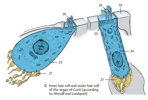
The
sensory cells (C) include the inner haircells (C23), which form only one row, andthe outer hair cells (C24);
the latter form three rows in the basal convolution of the cochlea, four rows
in the middle convolu-tion, and five rows in the upper convolution. All hair
cells have below their apical surface a dense terminal web of microfilaments (C25) through which the sensory hairs (C26) are firmly secured. The sensory
hairs are stiff, specialized microvilli (stereovilli) which are arranged in a
semicircle, usually in three rows of different lengths. Nerve fibers with
synapse – like contacts (C27)
ter-minate at the base of the hair cells.
Related Topics