Chapter: Biotechnology Applying the Genetic Revolution: Nanobiotechnology
Nanoparticles for Labeling
NANOPARTICLES
FOR LABELING
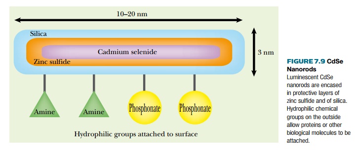
Consider luminescent CdSe
nanorods as an example of nanoparticles used for labeling (Fig. 7.9). These
nanorods can be used as fluorescent labels for molecular biology because they
absorb light from the UV to around 550 nm and emit strongly at 590 nm. They
were made—appropriately enough—in the lab of Thomas Nann, in Freiberg, Germany.
These nanorods measure approximately
3 nm in width by 10 to 20 nm in length. A core of luminous cadmium selenide
(CdSe) is surrounded by a shell of ZnS (zinc sulfide, wurtzite) to protect the
core against oxidation. Outside this is a layer of silica, which allows
coupling of phosphonates or amines to the exterior of the nanorod. These
hydrophilic groups make the nanorods water soluble. These outer chemical groups
also allow attachment of the nanorods to proteins.
The scaffold inside eukaryotic cells is built from cylindrical protein structures known as microtubules. These are often disassembled into monomers (known as tubulin) and reassembled in different locations. Nanorods can be used to follow this remodeling by attaching them to the tubulin monomers. On addition of guanosine triphosphate (GTP), assembly of microtubules is stimulated and the fluorescent nanorods can be seen aggregating into linear structures.
Why use a complex
multilayered nanostructure instead of a simple fluorescent dye?
(a) Although nanocrystals have narrow emission peaks, they
have broad absorption peaks (rather
than narrow ones like typical dyes). Consequently they do not bleach during
excitation and can therefore be used for continuous long-term irradiation and
monitoring.
(b) Nanocrystals have high brightness—the product of molar
absorptivity and quantum yield. (Molar absorptivity is the absorbance of
a one molar solution of pure solute at a given wavelength; the higher it is,
the more light is absorbed. The quantum
yield is the ratio of photons absorbed to photons emitted during
fluorescence.)
(c) The emission maximum of a nanocrystal depends on the
size and so can be set to any desired
wavelength by making crystals of the appropriate size (see later discussion).
Nanoparticles can also be
targeted to specific tissues, such as cancer cells, by adding appropriate
antibodies or receptor proteins to the nanoparticle surface. Fluorescent
nanoparticles are often known as quantum dots and are now commercially
available for a wide range of biological labeling. Although fluorescent dyes
can be attached to other molecules, nanoparticles are more versatile in this
regard. Quantum dots can be used to label DNA molecules as well as proteins.
Thus labeling of PCR primers with quantum dots results in fluorescently labeled
PCR products—a variant referred to as quantum
dot PCR.
A variety of materials have
been used to give better contrast enhancement in MRI. Nanoparticles containing
a variety of materials are beginning to see increasing use in this area. For
example, superparamagnetic iron oxide (SPIO) nanoparticles act as good MRI
contrast agents. Their magnetic properties vary with particle size. Larger
particles, of greater than 300 nm, are used for bowel, liver, and spleen.
Smaller particles, of 20 to 40 nm, have shown higher diagnostic accuracy for
detecting early tumors in lymph nodes than conventional materials.
Related Topics