Chapter: Biotechnology Applying the Genetic Revolution: Nanobiotechnology
Atomic Force Microscopy
ATOMIC
FORCE MICROSCOPY
Visualization at the
nanoscale is often performed using atomic force microscopy. As the name
indicates, this operates by measuring force, not by using a stream of particles
such as photons (as in light microscopy) or electrons (as in electron
microscopy).
Physicists sometimes compare
the operation of an AFM to an old-fashioned record player, which uses a needle
to scrape the surface of a record. Perhaps to a biologist, the difference
between a light microscope and AFM is like the difference between reading text
with the eyes and feeling Braille.
The atomic force microscope
was invented in 1985 by Gerd Binnig, Calvin Quate, and Christof Gerber. The AFM
uses a sharp probe that moves over the surface of the sample and which bends in
response to the force between the tip and the sample. The movement of the probe
performs a raster scan and the resulting topographical image is displayed
onscreen.
During scanning, the movement
of the tip or sample is performed by an extremely precise positioning device
and is made from piezoelectric ceramics. (These are materials that
change shape in response to an applied voltage.) It usually takes the form of a
tube scanner that is capable of sub-Ångstrom resolution in all three
directions.
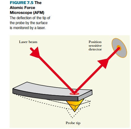
The AFM probe is a tip on the
end of a cantilever. As the cantilever bends because of the force on the tip,
its displacement is monitored by a laser, as shown in Fig. 7.5. The beam from
the laser is reflected onto a split photodiode. The difference between the A
and B signals measures the changes in the bending of the cantilever. For small
displacements, the displacement is proportional to the force applied. Hence the
force between the tip and the sample can be derived.

The distance between tip and
sample is adjusted so that it lies in the repulsive region of the
intermolecular force curve; that is, the AFM probe is repelled by its molecular
interaction with the surface. The repulsion gives a measure of surface
topography, and this is what is generally displayed, with color coding
indicating relative height. It is possible to scan a surface for topography and
then raise the AFM probe and rescan to detect electrostatic or magnetic forces.
These can then be plotted for comparison with the topography.
As with STM, it is possible
to use AFM to move single atoms, although this was only achieved in 2003.
Researchers at Osaka University in Japan removed a single silicon atom from a
surface and then replaced it.
Using AFM, it is possible to
visualize polymeric biological molecules such as DNA or cellulose and even to
see the individual monomers and, at high resolution, even the atoms of which
they are composed.
Related Topics