Chapter: Medical Physiology: Pregnancy and Lactation
Maturation and Fertilization of the Ovum
Maturation and Fertilization of the Ovum
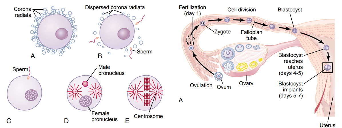
While still in the ovary, the ovum is in the primary oocyte stage. Shortly before it is released from the ovarian follicle, its nucleus divides by meiosis and a firstpolar body is expelled from the nucleus of the oocyte. The primary oocyte thenbecomes the secondary oocyte. In this process, each of the 23 pairs of chromo-somes loses one of its partners, which becomes incorporated in a polar body that is expelled. This leaves 23 unpaired chromosomes in the secondary oocyte. It is at this time that the ovum, still in the secondary oocyte stage, is ovulated into the abdominal cavity. Then, almost immediately, it enters the fimbriated end of one of the fallopian tubes.
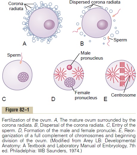
Entry of the Ovum into the Fallopian Tube (Oviduct). When ovulation occurs, the ovum,along with a hundred or more attached granulosa cells that constitute the corona radiata, is expelled directly into the peritoneal cavity and must thenenter one of the fallopian tubes to reach the cavity of the uterus. The fimbri-ated ends of each fallopian tube fall naturally around the ovaries. The inner sur-faces of the fimbriated tentacles are lined with ciliated epithelium, and the cilia are activated by estrogen from the ovaries, which causes the cilia to beat toward the opening, or ostium, of the involved fallopian tube. One can actually see a slow fluid current flowing toward the ostium. By this means, the ovum enters one of the fallopian tubes.
It seems likely that many ova might fail to enter the fallopian tubes. However, on the basis of conception studies, it is probable that as many as 98 per cent succeed in this task. Indeed, in some recorded cases, women with one ovary removed and the opposite fallopian tube removed have had several children with relative ease of conception, thus demonstrating that ova can even enter the opposite fallopian tube.
Fertilization of the Ovum. After the male ejaculates semen into the vagina duringintercourse, a few sperm are transported within 5 to 10 minutes upward from the vagina and through the uterus and fallopian tubes to the ampullae of the fallopian tubes near the ovarian ends of the tubes. This transport of the sperm is aided by contractions of the uterus and fallopian tubes stimulated by prostaglandins in the male seminal fluid and also by oxytocin released from the posterior pituitary gland of the female during her orgasm. Of the almost half a billion sperm deposited in the vagina, a few thousand succeed in reaching each ampulla.
Fertilization of the ovum normally takes place in the ampulla of one of the fallopian tubes soon after both the sperm and the ovum enter the ampulla. But before a sperm can enter the ovum, it must first pene-trate the multiple layers of granulosa cells attached to the outside of the ovum (the corona radiata) and then bind to and penetrate the zona pellucida surrounding the ovum itself.
Once a sperm has entered the ovum (which is still in the secondary oocyte stage of development), the oocyte divides again to form the mature ovumplus a second polar body that is expelled. The mature ovum still carries in its nucleus (now called the femalepronucleus) 23 chromosomes. One of these chromo-somes is the female chromosome, known as the Xchromosome.
`In the meantime, the fertilizing sperm has also changed. On entering the ovum, its head swells to form a male pronucleus, shown in Figure 82–1D. Later, the 23 unpaired chromosomes of the male pronucleus and the 23 unpaired chromosomes of the female pronu-cleus align themselves to re-form a complete comple-ment of 46 chromosomes (23 pairs) in the fertilizedovum (see Figure 82–1E).
What Determines the Sex of the Fetus That Is Created? Afterformation of the mature sperm, half of these carry in their genome an X chromosome (the female chromo-some) and half carry a Y chromosome (the male chromosome). Therefore, if an X chromosome from a sperm combines with an X chromosome from an ovum, giving an XX combination, a female child will be born. But if a Y chro-mosome from a sperm is paired with an X chromo-some from an ovum, giving an XY combination, a male child will be born.
Transport of the Fertilized Ovum in the Fallopian Tube
After fertilization has occurred, an additional 3 to 5 days is normally required for transport of the fertilized ovum through the remainder of the fallopian tube into the cavity of the uterus (Figure 82–2). This transport is effected mainly by a feeble fluid current in the tube resulting from epithelial secretion plus action of the ciliated epithelium that lines the tube; the cilia always beat toward the uterus. Weak contractions of the fallopian tube may also aid the ovum passage.
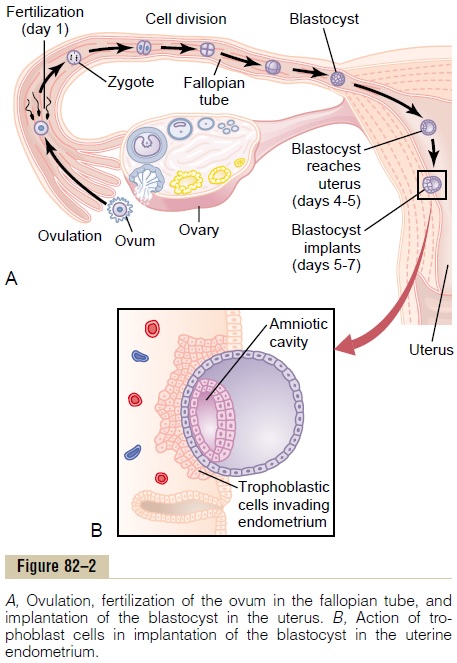
The fallopian tubes are lined with a rugged, cryptoid surface that impedes passage of the ovum despite the fluid current. Also, the isthmus of the fallopian tube (the last 2 centimeters before the tube enters the uterus) remains spastically contracted for about the first 3 days after ovulation. After this time, the rapidly increasing progesterone secreted by the ovarian corpus luteum first promotes increasing progesterone receptors on the fallopian tube smooth muscle cells; then the progesterone activates the receptors, exerting a tubular relaxing effect that allows entry of the ovum into the uterus.
This delayed transport of the fertilized ovum through the fallopian tube allows several stages of cell division to occur before the dividing ovum—now called a blastocyst, with about 100 cells—enters the uterus. During this time, the fallopian tube secretory cells produce large quantities of secretions used for the nutrition of the developing blastocyst.
Implantation of the Blastocyst in the Uterus
After reaching the uterus, the developing blastocyst usually remains in the uterine cavity an additional 1 to 3 days before it implants in the endometrium; thus, implantation ordinarily occurs on about the fifth to seventh day after ovulation. Before implantation, the blastocyst obtains its nutrition from the uterine endometrial secretions, called “uterine milk.”
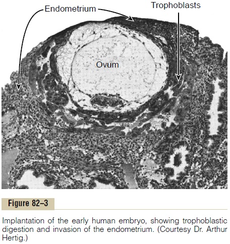
Implantation results from the action of trophoblastcells that develop over the surface of the blastocyst.These cells secrete proteolytic enzymes that digest and liquefy the adjacent cells of the uterine endometrium.
Some of the fluid and nutrients released are actively transported by the same trophoblast cells into the blas-tocyst, adding more sustenance for growth. Figure 82–3 shows an early implanted human blastocyst, with a small embryo. Once implantation has taken place, the trophoblast cells and other adjacent cells (from the blastocyst and the uterine endometrium) proliferate rapidly, forming the placenta and the various mem-branes of pregnancy.
Related Topics