Morphology, Antigenic Structure, Pathogenicity, Laboratory Diagnosis, Treatment and Preventions - Leptospira Interrogans | 12th Microbiology : Chapter 7 : Medical Bacteriology
Chapter: 12th Microbiology : Chapter 7 : Medical Bacteriology
Leptospira Interrogans
Leptospira Interrogans
Spirochaetes
of the genus Leptospira are actively
motile, delicate and possess numerous closely wound spirals with characteristic
hooked ends. Several Leptospires are saprophytes, while many are potential
pathogens of rodents, domestic animals and humans. The genus Leptospira
consists of two important species, which are Leptospira interrogans and Leptospira
biflexa.
Leptospira interrrogans is the causative agent of leptospirosis, a
zoonotic disease. The word Leptospira is derived from Latin word ‘Leptos’ =
fine or thin and ‘spira’ = Coil and interrogans = Question mark (The shape of
this spirochete accounts for its name)
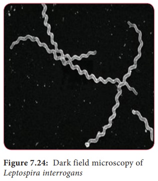
Morphology
• They
are spiral bacteria (5–20µm × 0.1µm) with numerous closely set coils. Their
ends are hooked and resemble umbrella handles.
• They are actively motile by rotatory movements.
• They cannot be seen under light microscope due to
its thinness, best observed by dark fieldmicroscopy (Figure 7.24), phase
contrast and electron microscope.
• They
stain poorly with aniline dyes, it may be stained with giemsa stain or silver
impregnation techniques.
Antigenic Structure
Leptospires
show considerable antigenic cross reaction.
a. Genus – Specific somatic antigen – It is present
in all members of the genus.
b. Surface
antigens – This antigen is used to classify Leptospira into serogroups and
serotypes.
Pathogenicity
Source of
infection: Contaminated water Route of entry: Through cuts or abrasions on skin
or mucosa
Incubation
period: 6–8 days
• Leptospira interrogans causes a zoonotic
disease named Leptospirosis. It is transmitted to humans by direct or indirect
contact with water, contaminated by urine of carrier animals (rat and dog).
• Leptospira enter the body through cuts or
abrasions on skin or through mucous membranes of the mouth, nose or
conjunctiva.
• After an
incubation period of 6–8 days. There is onset of febrile (related to fever)
illness with Leptospira in blood (Septicemic phase) which lasts for 3–7 days.
• The organisms disappear from the blood and
invades liver, kidney, spleen, meninges producing meningeal irritation such as
headache, vomiting.
• The pathogen persists in the internal organs and
most abundantly in the kidney. Severe Leptospirosis (Weil’s disease) is
associated with Fever, conjunctivitis (inflammation of conjunctiva),
albuminuria (presence of albumin in the urine), jaundice and hemorrhage. It is
a fatal illness with hepatorenal (Kidney failure with severe liver damage).
Clinical manifestations
• In severe cases, vomiting, headache, irregular
fever and intense infection of the eyes
• Jaundice, Albuminuria (The presence of protein
Albumin in the urine) and purpuric hemorrhages sometimes occur on skin and
mucosa.
Laboratory Diagnosis
The diagnosis
of Leptospirosis is made by the following ways
• Direct microscopy of blood or urine
• Isolation of pathogen by culture
• Serological tests.
Direct Microscopy
Blood: Leptospira can be observed in the blood by dark – filed microscope.
Blood examination is useful in first week as Leptospira disappear from blood
after 8 days.
Urine: Leptospira
may be present in urine in the 2nd
week of the disease and intermittently thereafterup to 6 weeks. Centrifuged
deposit of urine may be observed by Dark filed microscopy.
Culture:
Blood (1st week) and urine (2nd–6 week) can be cultured in Korthof ’s medium. Media are
incubated at 37°C for 2 days and then left at room temperature for 2 weeks.
Culturesare examined every third day for the presence of Leptospira under DFM
Serological tests
It is
very useful method of diagnosis two types of serological tests are used, which
are,
a. Screening tests: These
tests are genus – specific and done
using reactive genus specific antigen (non – pathogenic L. biflexapatoc I
strain).
Screening
test includes – CFT, ELISA, SEL, HAT indirect IF these tests are capable to
detect IgM and IgG leptospiral antibodies.
b. Serotype
specific tests: These tests identify
the infecting serovar by demonstrating specific antibodies..
i. Macroscopic agglutination test
ii. Microscopic agglutination test
Treatment and Preventions
Leptospira
are sensitive to penicillin and tetracycline
Preventive measures include rodent control,
disinfection of water.
Related Topics