Chapter: Biochemistry: Immunology
Immunity
Immunity
Disease spreads from one person to another is
called as infectious disease. Infectious diseases are caused by foreign
substances like fungi, bacteria, virus or parasite, when they enter in to the
human body. Though the disease by such pathogen affects the body for a shorter
duration, the person may survive after loosing functions of some of the organ
(eg. Poliomyelitis). Some times, when the infection or disease is severe the
person has to die. Yet many of the human beings are able to lead a normal life
because of the immune system. The immune system provides such freedom enjoyed
by an individual, in order to keep them free from diseases.
The immune system has the following functions,
·
Recognition
and defense against foreign substances (antigen) irrespective of the route of
entry.
·
Depending
upon the nature of the pathogen, appropriate immune reaction is mounted.
·
The
antigen induced Antibody combine specifically to that antigen (Specificity)
Immune system keep memory about the pathogens
and when the same pathogen reenters a better immune response is produced. This
forms the basis of vaccination.
·
Recognition
and destruction of the mutant cells that can become cancerous and this is known
as Immunosurveillance.
·
Normally,
Immune system does not produce antibodies against its own body tissues (self
antigens), called as Immune tolerance or
Self recognization.
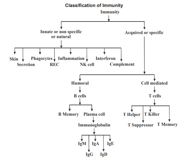
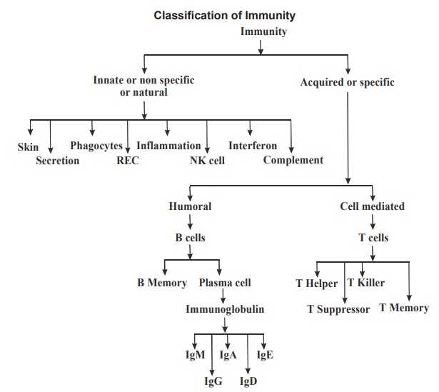
Depending on the
nature of response towards the pathogen, Immune system is broadly classified
into Natural and Acquired immunity. Immune system is classified as follows.
1. Natural Immunity
The non specific immunity present from birth is
known as innate immunity or natural immunity. It protects the
body against any foreign invaders and does not show any specificity. It is also
functionally matured in a new born. It does not become more efficient after
subsequent exposures to same organism.
Components (Cell types) involved in immunity
The cells of the immune system include
leukocytes, which are also known as white blood cells (WBC). They developed
from the bone marrow stem cells and give rise to two families of white blood
cells namely the Myeloid cells (named
after bone marrow) and the Lymphoid
cells, which take their name from the lymphatic system. Myeloid cells include Basophils, Eosinophils and Neutrophils.
The monocytes
give rise to macrophageswhen enter
into the tissue spacefrom blood circulation. Similarly, Basophilare transformed to mast
cells. Thelymphoid cells include T and B lymphocytes which get their maturation in different lymphoid organs.
B-cell maturation begins in the liver (fetal) and continues within thebone
marrow as maturation progresses (adult) and T cells complete their maturationin the thymus.
Mechanisms involved in Natural immunity Skin barrier
The skin covers and protects the body as a
barrier to prevent invading pathogens. Intact skin prevents the penetration of
most pathogens, by secreting lactic acid and fatty acids which lower the skin
pH.
Mechanical barriers
Mucous membranes form the external layer where
body is not covered with skin and it plays an important role in the prevention
of pathogen entrance by traping them. Movement of the mucociliary process in
the upper respiratory tract, the cilia in the eyelids act as escalators to
remove the pathogens.
Secretions
Sweat has antibacterial substances and tears
contain lysozyme. Mucous secretion in nose prevents the dust and microorganism
entry into the respiratory tract. Saliva contains lysozyme, thiocyante and
lactoferrin. The HCl acid secreted in the stomach kills the microbes.
Phagocytosis
The ingestion (endocytosis) and killing of
microorganisms by specialized cells called as phagocytes. Phagocytes are
polymorphonuclear leukocytes (eg.Neutrophils) and mononuclear cells (Monocytes
and Macrophages). Opsonization -The
process by which microbes are coated by a molecule called opsonin which aids
attachment of microbes to the phagocytic cells which facilitates phagocytosis.
Neutrophils constitutively express ligands and receptors (L-selectin) which
interact with reciprocal receptors and ligands on endothelial cells (P- and
E-selectin).The endothelial cells are located in the innermost layer of the
blood vessels. These interactions help the neutrophils to marginate and roll along
the endothelium. Neutrophil responds and move towards a group of molecules
called chemo-attractants (chemical mediators) and this process is called chemotaxis (chemical attraction). The
phagocytes make its way through intact capillary walls and into the surrounding
tissue by a process called diapedesis
(emigration of phagocytes into tissues). Chemo-attractants include complement
protein C5a, bacterial products, cytokines, lipid mediators from injured
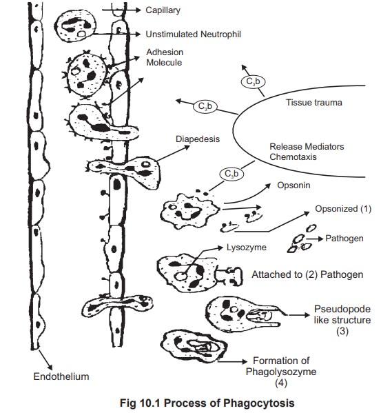
tissue. The various stages of Phagocytosis given below.
Stages of Phagocytosis (Fig. 10.1)
Opsonization (process by which microbes are
coated by a molecule called opsonin). Attachment to the pathogen (so that
pathogen movement can be restricted).
·
Formation
of Pseudopodia (hand like projections).
·
Encircling
of pathogen by pseudopodia leads to the formation of Phagosome.
·
Fusion
of Phagosome with lysozyme vesicle leads to the formation of phagolysosome.
Killing of Pathogen.
Killing by phagocytes
Neutrophil are able to kill the pathogen as they
posses certain chemicals in the form of granules and also the lysozyme enzyme.
Neutrophil invasion to an inflammed area is consider as the second line of
defence. Neutrophil has three types of granules namely Primary granules( contain serine proteases, lysozyme and
phospholipase A2) Secondary
granules ( include perforrin, elastase and collagenase) and Tertiarygranules ( contain gelatinase).
Apart from these granules the phagocytes alsoposses a variety of oxygen dependent killing mechanisms.
Phagocytes produce a respiratory burst, which produces superoxides and hydrogen
peroxide. Neutrophils contain an enzyme called as myeloperoxidase, which can convert superoxide into hypochlorite ion
which has a strong bactericidal activity.
Reticulo endothelial system (RES)
A diffuse system of cells that includes
monocytes and macrophages, which are phagocytic in nature. The role of
macrophage is consider as first order defence mechanism, as it engulf and kill
more pathogens efficiently. Macrophages also takes part in antigen
presentation. Apart from this, RES also involved in removing aged RBCs, denatured
protein, steroids,dyes and drugs.
The macrophages derive the name according to
their location.
Liver
- Kupffer cells
Brain Microglial cells
Kidney Mesangial cells
Spleen Splenic macrophages
Peritoneum
Peritoneal
macrophages.
Alveoli Alveolar macrophages.
Inflammation
A localized protective reaction produced in
tissue response to any irritation, injury or infection is called as
inflammation. This is characterized by pain, redness, swelling, and sometimes
loss of function. Usually, the name of the tissue, organ and the region which
develops inflammation is suffixed with ‘itis’
for example conjunctivitis, gastritis and pharyngitis respectively. The
inflammatory response helps to mobilize the nonspecific defense forces to the
tissue space where pathogen is present. The damaged cells release chemical
mediators such as histamine from the mast cells, which dilate the near by blood
vessels. The complement system gets activated and attracts phagocytes. The
plasma leaking from the dilated blood vessel contains clotting system of
proteins. They get activated due to the tissue damage and this process leads to
“walling off” the area and this helps to prevent spreading of the infectious
material.
Natural Killer Cells
Among the immune cells, natural killer cells (NK
cells) are the most aggressive. They are first line of defense against infected
and cancerous cells. They are lymphocytes (Large granular lymphocytes, LGL)
with no immunological memory and are part of the innate immune system. It
attaches to the target and releases a lethal burst of chemicals called as
perforins that penetrate the cell wall. Fluids begin to leak in and out and
eventually the cell explodes.
Interferon
Interferons are proteins produced by body cells
when they are invaded by viruses, is released into the bloodstream or
intercellular fluid, in order to induce healthy cells to manufacture an enzyme
that block viral replication.
Complement System
It is a group of proenzymes. They circulate in
serum in inactive form. The complement system is the part of innate immune
system plays an important defense against microorganisms, especially
gram-negative bacteria. The complement system consists of a set of over twenty
serum proteins which are getting activated as follows.
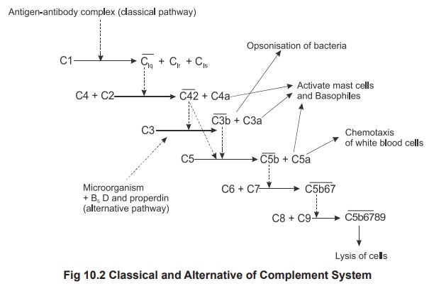
The complement cascade consists of two separate
pathways that converge in a final common pathway (Fig.2). The pathways include
the classic pathway (C1qrs, C2, C4),
the alternative pathway (C3, factor
B, properdin) and these two pathways converge at the component C3. The terminal
complement pathway consists of all proteins activated after C3. The most
notable are C5-C9 group of proteins collectively
known as the membrane
attack complex (MAC). The MAC exerts powerful killing activity by creating
perforations in cellular membranes. Activated C3b opsonizes bacteria and C5a
function as chemotactic agent.
Antigen presenting cells (APC)
B cell,dendritic cells (lymphnodes), Langerhans
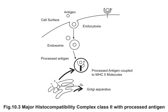
cells (from skin) and macrophages are called as antigen presenting cells. All
these cells, process the antigen and express the antigen over the surface of
its cell membrane along with a molecule called as Major Histo Compatibility
Complex (MHC) class II molecule.
Major Histocompatability complex
A set of cell surface glycoproteins are called
as the Major Histocompatibility Complex or MHC molecules. Generally, they take
part in differentiating self and non self antigens and the presentation of
processed foreign antigen to activate the T cells. There are two classes of MHC
proteins, MHC class I and MHC class II. MHC class I molecule is expressed on
the cell surface of all nucleated cells of the body. MHC class I molecules with
processed antigen are expressed on the surface of the infected cells, which
present the processed antigen to cytotoxic T cells (CD8). MHC class II molecule
are expressed on APC cell surface which present the processed antigen to Helper
T cells (CD4 cells).
2. Acquired immunity
Producing specific cells and molecules which are
directed against the foreign invaders. It has the special ability to keep
memory of first time exposure of an antigen (primary immune response) and
mounts better response when there is second time exposure of same antigen
(secondary immune response). This ability of immune response forms the basis
for the immunization or vaccination.
Acquired immunity is classified into humoral
immunity and cell mediated immunity. Both humoral and cellular immune responses
are evoked during antigen exposure.
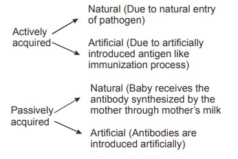
The acquired immunity can be either active or
passive.
3. Humoral immunity
The humoral immune response begins with the
recognition of antigen. Though the classification separates the cell mediated
and humoral immunity with different cell types they do interact to bring an
effective immune response. Specific T-cells are stimulated to produce
lymphokines that are responsible for the antigen-induced B-cells proliferation
and differentiation.
This is for the T depended antigens. However
some of the macro antigenic molecules can directly stimulate the B cells
directly. Through a process of clonal selection specific B-cells are
stimulated, the activated B-cell first develops into a B-lymphoblast, becoming
much larger and shedding all surface immunoglobulin. This terminal
differentiation stage is responsible for production of primarily IgM antibody
during the primary immune response. Few newly differentiated B-cells remain as
long-lived “memory cells” without secreting antibodies. Upon subsequent
encounter with antigen, these cells respond very quickly to produce large
amounts of IgG, IgA or IgE antibody, generating the better secondary immune
response.
Pathogen
or foreign protein + Macrophage / dentritic cells →
processed antigen
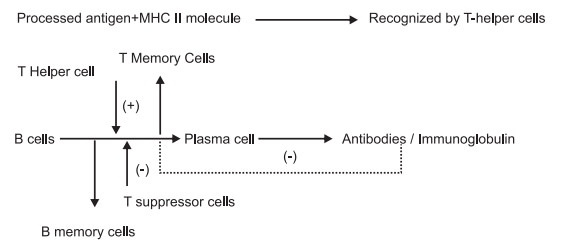
The initial differentiation step that ultimately
leads to the mature B-cell involves DNA rearrangements in heavy chain variable
(V) region as well as similar rearrangements within the light chain genes to
synthesis immunoglobulin. These stages are, of course, initiated upon encounter
with antigen and activation by T-helper cell to secrete lymphokines. The
activated B-cell first develops into a B-lymphoblast, becoming much larger. IgM antibody is formed during the ‘primaryimmune response.’ Instead,
these cells undergo secondary DNA rearrangements tomodify the constant region
and forms IgG, IgA or IgE antibodies
during secondaryimmune response. The
suppressor T-cells suppresses the immune response oncean adequate amount of
antibody formed. Another way of suppression occurs by the produced antibody
itself and known as, “antigen blocking”. When high doses of antibody interact
with the entire antigen’s epitopes thereby inhibits interactions with B-cell
receptors.
4. Cell mediated immunity
T cells are responsible for cell-mediated immunity. T cells are initially formed in the bone
marrow and get its maturation and differentiation in the thymusgland. After maturation T cells migrate to secondary lymphoid organ. T cells
areclassified according to their functions and cell-surface marker called CDs (clusters ofdifferentiation). They are
functionally classified as T helper, T suppressor, T memoryand T killer cells. T cells are associated with certain
types of allergic reactions called Delayed
hypersensitivity and also in transplanted organ rejection.
The Major
Histocompatibility Complex (MHC) are unique to each individual and indicate
self-molecules and always these
molecules are given as reference when ever an antigen is presented and this
helps the immune system to differentiate the self from non-self. (fig. 3) Helper T (TH) cells (also
known as CD4 cells) activate B cells
to produce antibodies against T-dependent antigens (usually protein in
composition). TH cells recognize and bind to an antigen in association with an MHC II (Fig. 3) on the surface of an
antigen presenting cells (APC) and
the APC cell secrete the cytokineIL-1 and induce the THcell
to secrete the cytokine IL-2. Only THcells
that have been stimulated by an antigen have
receptors for IL-2 and thus these THcells are specificfor only that
stimulatory antigen. Production of IL-2 and other cytokines by these TH
cells stimulates the cell-mediated (e.g., TC cells) and humoral (B
cells- Plasma cell) immune responses. In Acquired Immuno Deficiency Syndrome
(AIDS),the Human Immuno deficiency virus (HIV) affect the T helper cells. Suppressor (TS) cells appear
to regulate the immune response once the antibody formation reached the
adequate levels. Cytotoxic T cells (CD8) identify the viral infected cells and
inject the molecule called Perforin to lyse the viral infected cells. Some of
the activated T cells become Tmemory
cells.
Role of lymphokines
Lymphokines are the cytokines secreted by the
lymphocytes and these are small molecules released due to a stimulus and help
to send the signal between cells. The term interleukin (IL) is also often
referring to the cytokine produced by leukocytes. There is considerable overlap
between the actions of the individual lymphokines, so that many of the above
effects are shared between TNFa, IL-2 to IL-12. In addition, these
proinflammatory cytokines activate the immune system, mobilizing neutrophils
from bone marrow, causing dendritic cells to migrate to lymph nodes, and also
initiating changes in adipocyte and muscle metabolism and also responsible for
inducing fever.
Related Topics