Chapter: 12th Zoology : Chapter 2 : Human Reproduction
Human reproductive system
Human
reproductive system
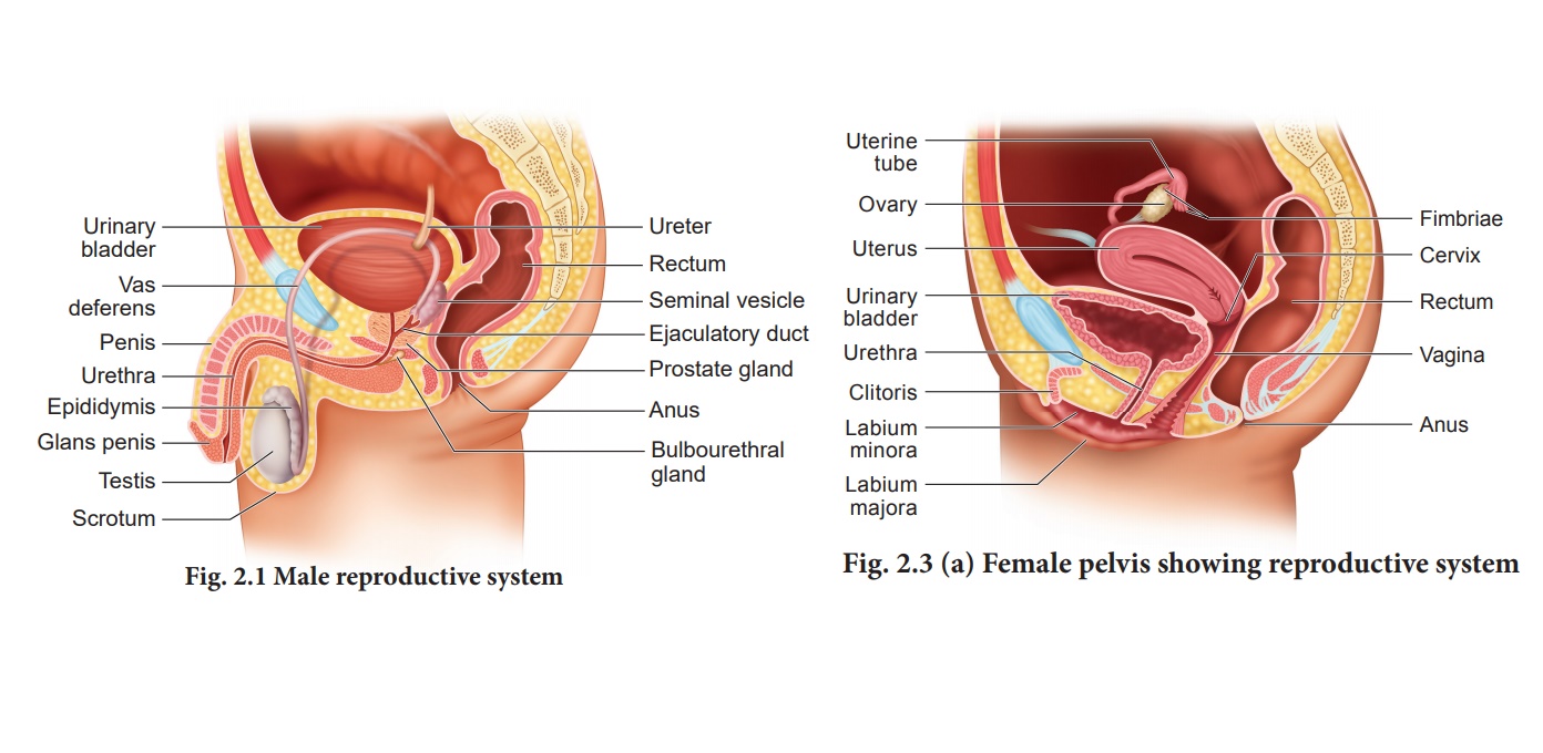
The male reproductive
system comprises of a pair of testes, accessory ducts, glands and external
genitalia (Fig. 2.1).
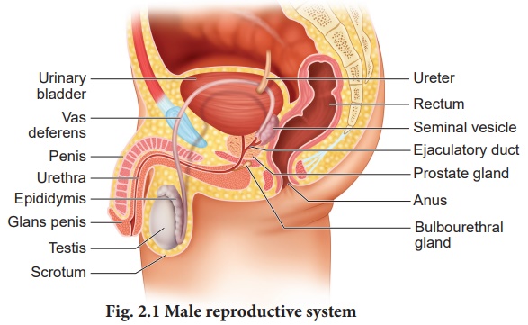
Testes are the primary
male sex organs. They are a pair of ovoid bodies lying in the scrotum (Fig.2.2
a). The scrotum is a sac of skin that hangs outside the abdominal cavity.
Since viable sperms cannot be produced at normal body temperature, the scrotum
is placed outside the abdominal cavity to provide a temperature 2-3oC lower
than the normal internal body temperature. Thus, the scrotum acts as a thermoregulator
for spermatogenesis.
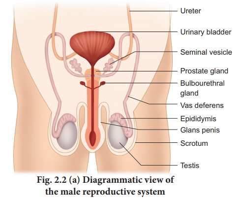
Each testis is covered
by an outermost fibrous tunica albuginea and is divided by septa into
about 200 - 250 lobules each containing 2-4 highly coiled testicular
tubules or seminiferous tubules. These highly convoluted tubules which form 80
percent of the testicular substance are the sites for sperm production.
The stratified
epithelium of the seminiferous tubule is made of two types of cells namely
sertoli cells or nurse cells and spermatogonic cells or male germ cells. Sertoli
cells are elongated and pyramidal and provide nourishment to the sperms
till maturation. They also secrete inhibin, a hormone which is involved
in the negative feedback control of sperm production. Spermatogonic cells
divide meiotically and differentiate to produce spermatozoa.
Interstitial cells or Leydig cells are embedded in the
soft connective tissue surrounding the seminiferous tubules.These cells are
endocrine in nature and secrete
androgens namely the testosterone hormone which
initiates the process of spermatogenesis. These cells are endocrine in nature
and are characteristic features of the testes of mammals. Other immunologically
competent cells are also present.
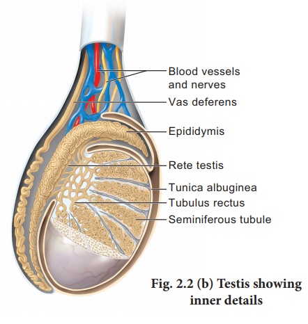
The accessory ducts
associated with the male reproductive system include rete testis, vasa
efferentia, epididymis and vas deferens (Fig.2.2 b). The
seminiferous tubules of each lobule converge to form a tubulus rectus
that conveys the sperms into the rete testis. The rete testis is a tubular
network on the posterior side of the testis. The sperms leave the rete testis
and enter the epididymis through the vasa efferentia. The epididymis is a
single highly coiled tube that temporarily stores the spermatozoa and they
undergo physiological maturation and acquire increased motility and fertilizing
capacity. The epididymis leads to the vas deferens and joins the duct of the
seminal vesicle to form the ejaculatory duct which passes through the prostate
and opens into the urethra. The urethra is the terminal portion of the male
reproductive system and is used to convey both urine and semen at different
times. It originates from the urinary bladder and extends through the penis by
an external opening called urethral meatus.
The accessory glands of the male reproductive system include the paired seminal vesicles and bulbourethral glands also called Cowper’s gland and a single prostate gland. The seminal vesicles secrete an alkaline fluid called seminal plasma containing fructose sugar, ascorbic acid, prostaglandins and a coagulating enzyme called vesiculase which enhances sperm motility.
The bulbourethral glands are inferior to the prostate and
their secretions also help in the lubrication of the penis. The prostate
encircles the urethra and is just below the urinary bladder and secretes a
slightly acidic fluid that contains citrate, several enzymes and prostate
specific antigens. Semen or seminal fluid is a milky white fluid which
contains sperms and the seminal plasma (secreted from the seminal vesicles,
prostate gland and the bulbourethal glands). The seminal fluid acts as a
transport medium, provides nutrients, contains chemicals that protect and
activate the sperms and also facilitate their movement.
The penis is the male
external genitalia functioning as a copulatory organ. It is made of a special
tissue that helps in the erection of penis to facilitate insemination. The
enlarged end of the penis called glans penis is covered by a loose fold of skin
called foreskin or prepuce.
The female reproductive
system is far more complex than the male because in addition to gamete
formation, it has to nurture the developing foetus. The female reproductive
system consists of a pair of ovaries along with a pair of oviducts, uterus,
cervix, vagina and the external genitalia located in the pelvic
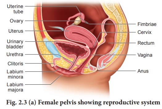
region (Fig. 2.3 a).
These parts along with the mammary glands are integrated structurally and
functionally to support the process of ovulation, fertilisation, pregnancy,
child birth and child care.
Ovaries are the primary
female sex organs that produce the female gamete, ovum. The ovaries are located
one on each side of the lower abdomen. The ovary is an elliptical structure
about 2-4 cm long. Each ovary is covered by a thin cuboidal epithelium called
the germinal epithelium which encloses the ovarian stroma. The stroma is
differentiated as the outer cortex and inner medulla. Below the germinal
epithelium is a dense connective tissue, the tunica albuginea.
The cortex appears dense
and granular due to the presence of ovarian follicles in various stages of
development. The medulla is a loose connective tissue with abundant blood
vessels, lymphatic vessels and nerve fibres. The ovary remains attached to the
pelvic wall and the uterus by an ovarian ligament called mesovarium.
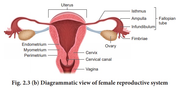
The fallopian tubes (uterine tubes or oviducts), uterus and vagina constitute the female accessory organs (Fig. 2.3 b). Each fallopian tube extends from the periphery of each ovary to the uterus. The proximal part of the fallopian tube bears a funnel shaped infundibulum. The edges of the infundibulum have many finger like projections called fimbriae which help in collection of the ovum after ovulation.
The infundibulum leads
to a wider central portion called ampulla. The last part of the oviduct is the
isthmus which is short and thick walled connecting the ampulla and infundibulum
to the uterus.
The uterus or womb is a
hollow, thick-walled, muscular, highly vascular and inverted pea shaped
structure lying in the pelvic cavity between the urinary bladder and rectum.
The major portion of the uterus is the body and the rounded region superior to
it, is the fundus. The uterus opens into the vagina through a narrow cervix.
The cavity of the cervix called the cervical canal communicates with the vagina
through the external orifice and with the uterus through the internal orifice.
The cervical canal along with vagina forms the birth canal.
The wall of the uterus
has three layers of tissues. The outermost thin membranous serous layer called
the perimetrium, the middle thick muscular layer called myometrium and the
inner glandular layer called endometrium. The endometrium undergoes cyclic
changes during the menstrual cycle while myometrium exhibits strong
contractions during parturition.
Vagina is a large
fibromuscular tube that extends from the cervix to the exterior. It isthe
female organ of copulation. The female reproductive structures that lie
external to the vagina are called as the external genitalia or vulva comprising
of labia majora, labia minora,hymen and clitoris.
The Bartholin’s
glands(also called greater vestibular glands) are located posterior to the left
and rightof the opening of the vagina.They secrete mucus to lubricate the
vagina and are homologous to the bulbourethral glands of the male. The Skene’s
glands are located on the anterior wall of the vagina and around the lower end
of the urethra. They secrete a lubricating fluid and are homologous to the
prostate gland of the males.
The external opening of
the vagina is partially closed by a thin ring of tissue called the hymen. The
hymen is often torn during the first coitus (physical union). However in some
women it remains intact. It can be stretched or torn due to a sudden fall or
jolt and also during strenuous physical activities such as cycling, horseback
riding, etc., and therefore cannot be considered as an indicator of a woman’s
virginity.
The mammary glands are
modified sweat glands present in both sexes. It is rudimentary in the males and
functional in the females. A pair of mammary glands is located in the thoracic
region. It contains glandular tissue and variable quantities of fat with a
median nipple surrounded by a pigmented area called the areola. Several
sebaceous glands called the areolar glands are found on the surface and they
reduce cracking of the skin of the nipple. Internally each mammary gland
consists of 2-25 lobes, separated by fat and connective tissues (Fig. 2.4).
Each lobe is made up of lobules which contain acini or alveoli lined by
epithelial cells. Cells of the alveoli secrete milk. The alveoli open into mammary
tubules. The tubules of each lobe join to form
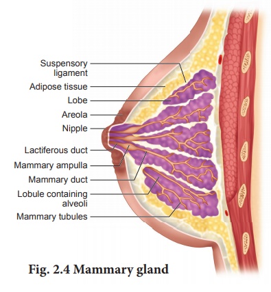
Several
mammary ducts join to form a wider mammary ampulla which is connected to the
lactiferous duct in the nipple. Under the nipple, each lactiferous duct expands
to form the lactiferous sinus which serves as a reservoir of milk. Each
lactiferous duct opens separately by a minute pore on the surface of the
nipple.
Normal development of the breast begins at puberty and progresses with changes during each menstrual cycle. In non-pregnant women, the glandular structure is largely underdeveloped and the breast size is largely due to amount of fat deposits.The size of the breast does not have an influence on the efficiency of lactation.
Related Topics