Chapter: Medical Surgical Nursing: Assessment and Management of Patients With Hearing and Balance Disorders
Function of the Ears
FUNCTION OF THE EARS
Hearing
Hearing
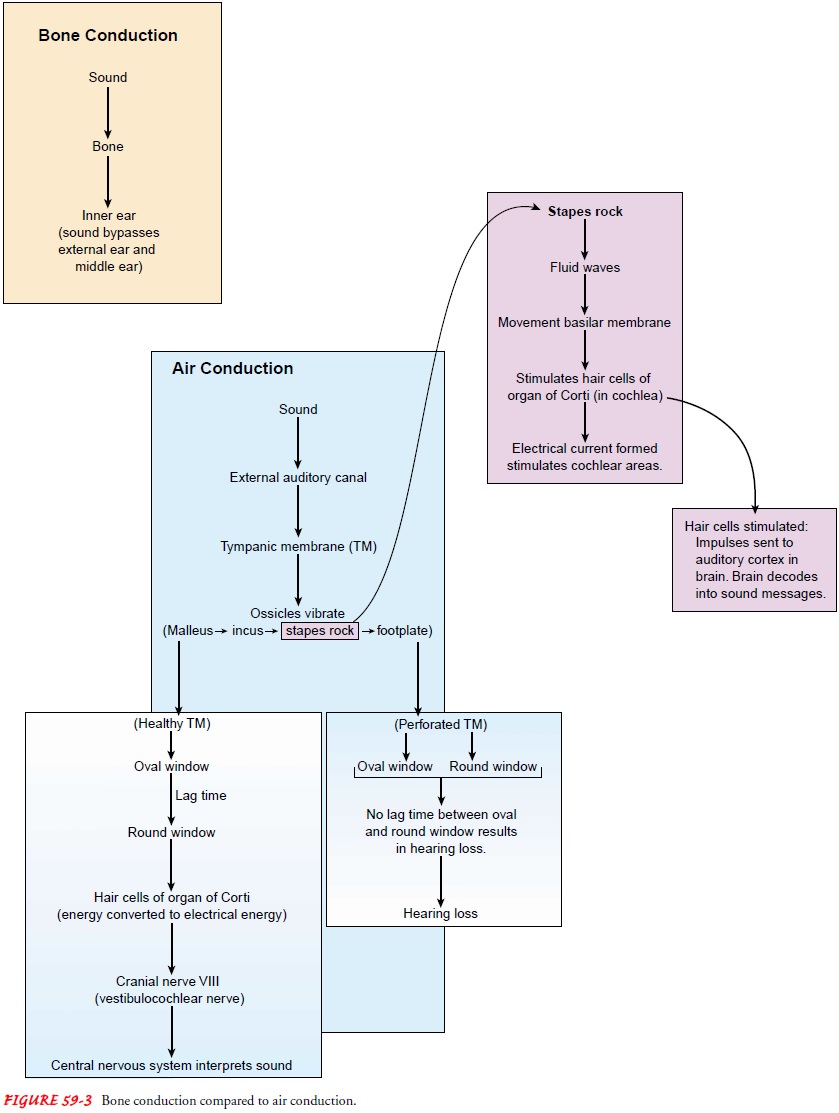
is conducted over two pathways: air and bone. Sounds transmitted by air
conduction travel over the air-filled external and middle ear through vibration
of the tympanic membrane and ossicles.Sounds transmitted by bone conduction
travel directly through bone to the inner ear, bypassing the tympanic membrane
and ossicles. Normally, air conduction is the more efficient path-way. However,
defects in the tympanic membrane or interrup-tion of the ossicular chain
disrupt normal air conduction, which results in a loss of the sound-to-pressure
ratio and subsequently in a conductive hearing loss.
SOUND CONDUCTION AND TRANSMISSION
Sound
enters the ear through the external auditory canal and causes the tympanic
membrane to vibrate. These vibrations transmit sound through the lever action
of the ossicles to the oval window as mechanical energy. This mechanical energy
is then transmitted through the inner ear fluids to the cochlea, stimulating
the hair cells, and is subsequently converted to electrical energy. The elec-trical
energy travels through the vestibulocochlear nerve to the cen-tral nervous
system, where it is analyzed and interpreted in its final form as sound.
Vibrations transmitted by the tympanic membrane to
the os-sicles of the middle ear are transferred to the cochlea, lodged in the
labyrinth of the inner ear. The stapes rocks, causing vibrations (ie, waves) in
fluids contained in the inner ear. These fluid waves cause movement of the
basilar membrane to occur that then stim-ulates the hair cells of the organ of
Corti in the cochlea to move in a wavelike manner. The movements of the
tympanic mem-brane set up electrical currents that stimulate the various areas
of the cochlea. The hair cells set up neural impulses that are encoded and then
transferred to the auditory cortex in the brain, where they are decoded into a
sound message.
The
footplate of the stapes receives impulses transmitted by the incus and the
malleus from the tympanic membrane. The round window, which opens on the
opposite side of the cochlear duct, is protected from sound waves by the intact
tympanic mem-brane, permitting motion of the inner ear fluids by sound wave
stimulation. For example, in the normally intact tympanic mem-brane, sound
waves stimulate the oval window first, and a lag oc-curs before the terminal
effect of the stimulus reaches the round window. This lag phase is changed,
however, when a perforation of the tympanic membrane is large enough to allow
sound waves to impinge on the oval and round windows simultaneously. This effect
cancels the lag and prevents the maximal effect of inner ear fluid motility and
its subsequent effect in stimulating the hair cells in the organ of Corti. The
result is a reduction in hearing ability (Fig. 59-3).

Balance and Equilibrium
Body balance is maintained by the cooperation of
the muscles and joints of the body (ie, proprioceptive system), the eyes (ie,
visual system), and the labyrinth (ie, vestibular system). These areas send
their information about equilibrium, or balance, to the brain (ie, cerebellar
system) for coordination and perception in the cerebral cortex. The brain
obtains its blood supply from the heart and ar-terial system. A problem in any
of these areas, such as arterioscle-rosis or impaired vision, can cause a
balance disturbance. The vestibular apparatus of the inner ear provides
feedback regarding the movements and the position of the head and body in
space.
Related Topics