Chapter: Medical Surgical Nursing: Assessment and Management of Patients With Hearing and Balance Disorders
Anatomy of the External Ear

ANATOMY OF THE EXTERNAL EAR
The
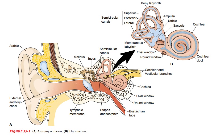
external ear, housed in the temporal bone, includes the au-ricle (i.e., pinna)
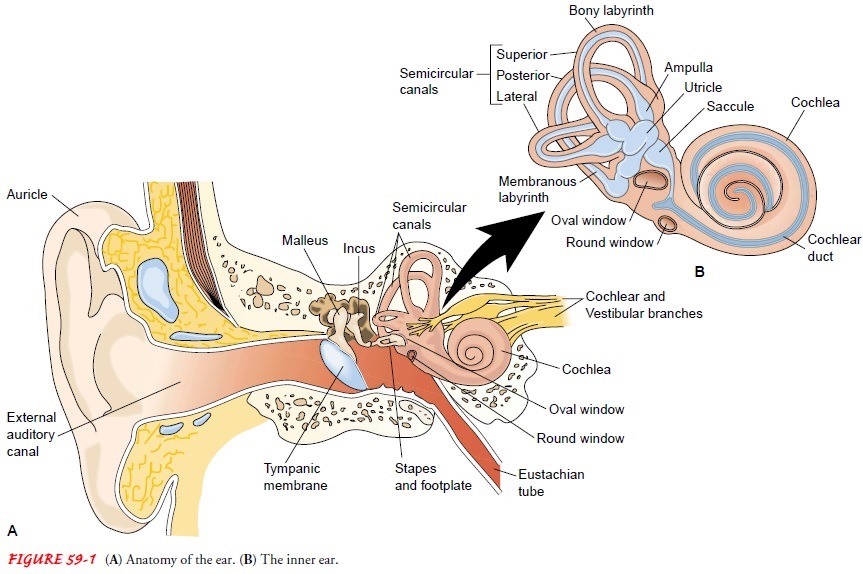
and the external auditory canal (Fig. 59-1). The external ear is separated from
the middle ear by a disklike struc-ture called the tympanic membrane (ie,
eardrum).

Auricle
The
auricle, attached to the side of the head by skin, is composed mainly of
cartilage, except for the fat and subcutaneous tissue in the earlobe. The
auricle collects the sound waves and directs vi-brations into the external
auditory canal.
External Auditory Canal
The
external auditory canal is approximately 2.5 cm long. The lateral third is an
elastic cartilaginous and dense fibrous frame-work to which thin skin is
attached. The medial two thirds is bone lined with thin skin. The external
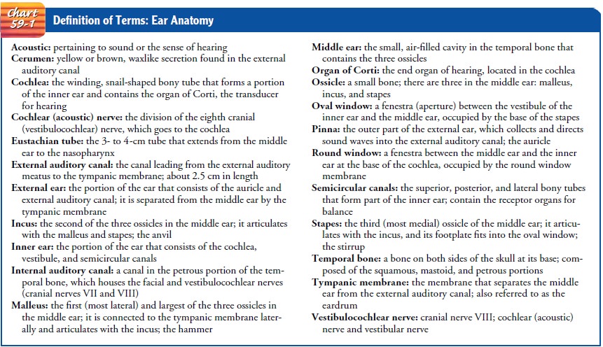
auditory canal ends at the tympanic membrane (Chart 59-1).

The
skin of the canal contains hair, sebaceous glands, and ceru-minous glands,
which secrete a brown, waxlike substance called cerumen (ie, ear wax). The
ear’s self-cleaning mechanism moves old skin cells and cerumen to the outer
part of the ear.
Just
anterior to the external auditory canal is the temporo-mandibular joint. The
head of the mandible can be felt by plac-ing a fingertip in the external
auditory canal while the patient opens and closes the mouth.
Related Topics