Chapter: Medical Surgical Nursing: Assessment and Management of Patients With Hearing and Balance Disorders
Anatomy of the Inner Ear

ANATOMY OF THE INNER EAR
The
inner ear is housed deep within the temporal bone. The organs for hearing (ie,
cochlea) and balance (ie, semicircular canals), as well as cranial nerves VII
(ie, facial nerve) and VIII (ie, vestibulocochlear nerve), are all part of this
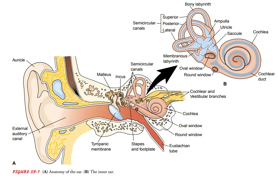
complex anatomy (see Fig. 59-1). The cochlea and semicircular canals are housed
in the bony labyrinth. The bony labyrinth surrounds and protects the membranous
labyrinth, which is bathed in a fluid called perilymph.

Membranous Labyrinth
The
membranous labyrinth is composed of the utricle, the sac-cule, the cochlear
duct, the semicircular canals, and the organ of Corti. The membranous labyrinth
contains a fluid called endo-lymph. The three semicircular canals—posterior,
superior, and lateral, which lie at 90-degree angles to one another—contain
sensory receptor organs, arranged to detect rotational movement. These receptor
end organs are stimulated by changes in the rate or direction of an
individual’s movement. The utricle and saccule are involved with linear movements.
Organ of Corti
The organ of Corti is located in the cochlea, a snail-shaped, bony tube about 3.5 cm long with two and one-half spiral turns. Mem-branes separate the cochlear duct (ie, scala media) from the scala vestibuli, and the scala tympani from the basilar membrane. The organ of Corti is located on the basilar membrane stretching from the base to the apex of the cochlea. As sound vibrations enter the perilymph at the oval window and travel along the scala vestibuli, they pass through the scala tympani, enter the cochlear duct, and cause movement of the basilar membrane. The organ of Corti, also called the end organ for hearing, transforms mechan-ical energy into neural activity and separates sounds into differ-ent frequencies. This electrochemical impulse travels through the acoustic nerve to the temporal cortex of the brain to be inter-preted as meaningful sound. In the internal auditory canal, the cochlear (acoustic) nerve, arising from the cochlea, joins the ves-tibular nerve, arising from the semicircular canals, utricle, and saccule, to become the vestibulocochlear nerve (cranial nerve VIII). This canal also houses the facial nerve and the blood supply from the ear to the brain.
Related Topics